Tagging with Fluorescent Proteins Allows the Visualization of Specific Proteins in Live Cells
The jellyfish Aequorea victoria expresses a naturally fluorescent protein, called green fluorescent protein (GFP, ~27 kDa). GFP contains a serine, tyrosine, and glycine sequence whose side chains spontaneously cyclize to form a green-fluorescing fluorochrome when illuminated with blue light. Using recombinant DNA technology, it is possible to make a DNA construct in which the coding sequence of GFP is fused to the coding sequence of a protein of interest. When this construct is introduced into and expressed in cells, a GFP- “tagged” protein is made in which the protein of interest is covalently linked to GFP as part of the same polypeptide. Although GFP is a medium-sized protein, the function of the protein of interest is often not changed by fusing it to GFP. This technique allows one to visualize GFP—and hence the protein of interest. One can not only see the location of the GFP-tagged protein immediately, but can also view its distribution in a live cell over time and thereby assess its dynamics or track its localization following various cell treatments. The simple idea of tagging specific proteins with GFP has revolutionized cell biology and has led to the development of many different fluorescent proteins (Figure 4-16). Use of this colorful variety of fluorescent proteins allows one to visualize two or more proteins simultaneously if they are each tagged with a different-colored fluorescent protein. We describe additional techniques that exploit fluorescent proteins in the following sections.
Page 147
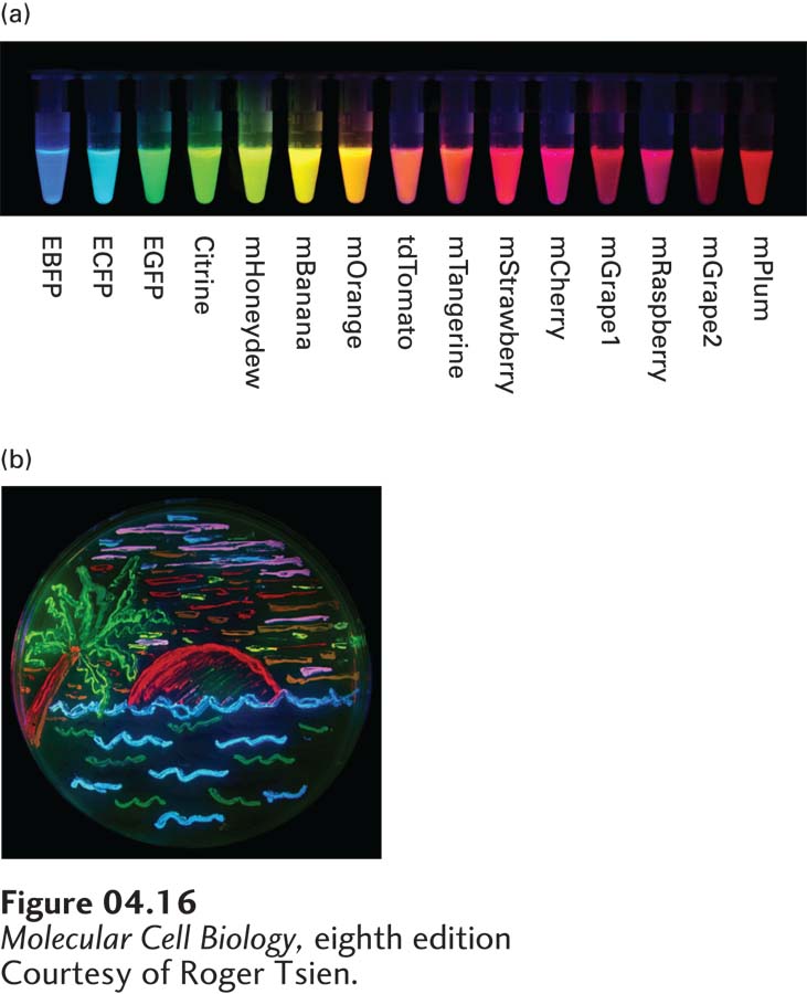
[Courtesy of Roger Tsien.]
FIGURE 4-16Many different colors of fluorescent proteins are now available. (a) Tubes show the emission colors and names of many different fluorescent proteins. (b) An agar dish is illuminated to show growing bacteria expressing several different-colored fluorescent proteins.