Two-Photon Excitation Microscopy Allows Imaging Deep into Tissue Samples
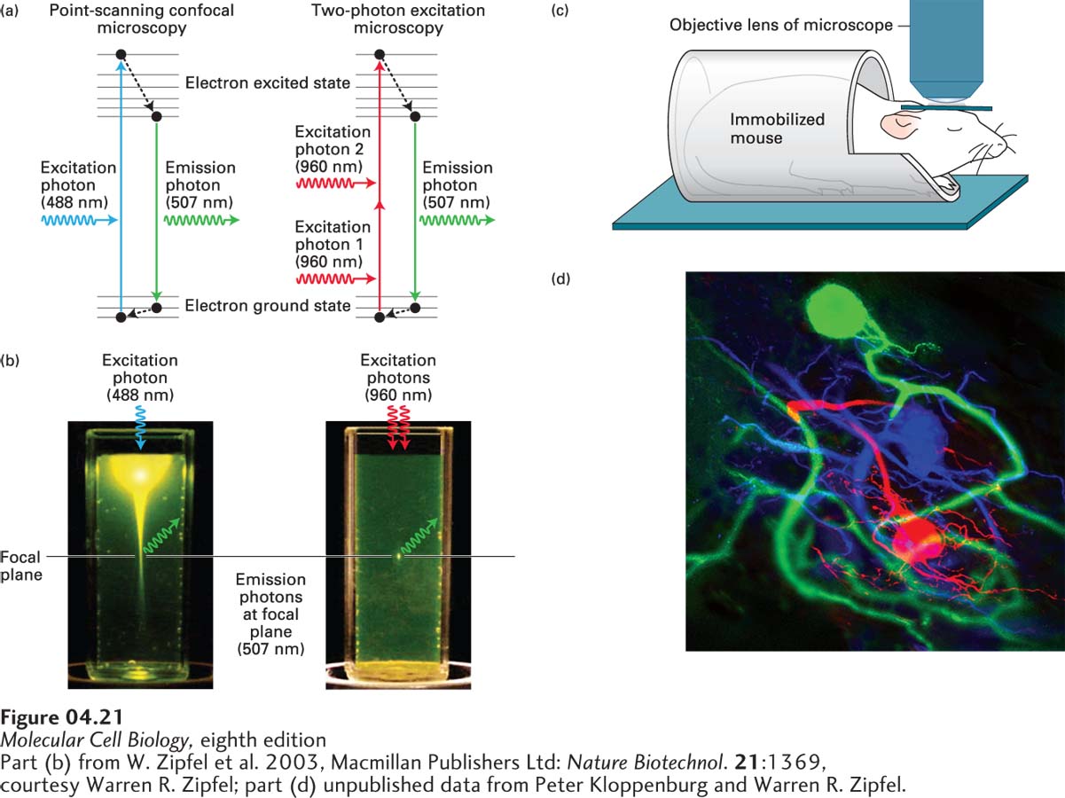
We have just seen how point-scanning confocal microscopy can help reduce fluorescence from out-of-focus planes. To achieve this, it focuses a cone of laser light on a spot that scans across the focal plane. Regions above and below the focal plane are also illuminated by this cone of light, generating out-of-focus signal that must be removed by collecting the light through a pinhole. This intense cone of light can also lead to photobleaching (rendering the fluorescent protein inactive) or damage to the sample by phototoxicity. If the sample is very thin, these are not significant problems, but as the sample gets thicker, they become more relevant. To circumvent these problems, use was made of the finding that a fluorochrome can be excited either by a single photon—for example, at 488 nm—or by two photons of half the energy at 960 nm, either of which will generate the same emission wavelength (Figure 4-21a). Thus if a 960-nm cone of laser light is focused on a spot in one plane so that only at the focal point is there sufficient density of photons to excite the fluorochrome (Figure 4-21b), no out-of-focus signal will be obtained and less photobleaching or phototoxicity will occur. Because only fluorochromes in the focal plane are excited, two-photon microscopy can be used to explore much thicker samples, and there is no need for a pinhole to exclude out-of-focus light. However, very high laser intensity is required for two-photon excitation microscopy, as the two photons must arrive within about a femtosecond of each other, which is achieved using rapid laser pulses. If individual cells in an animal express different color variants of fluorescent proteins, two-photon microscopy can be used to observe events in living animals (called intravital imaging) within about 1 mm of the surface (Figure 4-21c, d).
[Part (b) from W. Zipfel et al. 2003, Macmillan Publishers Ltd: Nature Biotechnol.21:1369, courtesy Warren R. Zipfel; part (d) unpublished data from Peter Kloppenburg and Warren R. Zipfel.]
FIGURE 4-21Two-photon excitation microscopy restricts illumination to the focal plane to allow deep penetration for intravital imaging. (a) A diagram illustrating the different excitation methods used for conventional point-scanning confocal microscopy and for two-photon excitation microscopy. In the conventional system, absorption of a single photon of the appropriate wavelength (here at 488 nm, shown by the blue arrow) results in an electron jumping to the excited state. After undergoing vibrational relaxation (black dashed arrow), the electron falls back to the ground state with emission of one photon at a longer (lower-energy) wavelength, in this case 507 nm (green arrow). In two-photon excitation, when two photons of the appropriate wavelength (shown here at 960 nm, red arrows) arrive almost instantaneously, they can both be absorbed and induce the electron to jump to the excited state. As in the previous case, the electron undergoes some vibrational relaxation (black dashed arrow) and falls back to the ground state with the emission of a photon (507 nm). (b) A cuvette of fluorescent material is illuminated with 488-nm light (left), as in conventional confocal microscopy, or with intense 960-nm light, as in two-photon microscopy. Notice that the conventional system produces a bright cone of excitation outside the focal plane, whereas two-photon excitation illuminates just one spot in the focal plane. (c) Because two-photon microscopy does not excite fluorochromes outside the plane of focus, it can be used to observe cells up to 1 mm deep within a living animal (“intravital imaging”). To image a living animal, it has to be immobilized on the microscope stage and access given for the objective lens to come close to the region being imaged. (d) An example of intravital imaging in which labeled neurons in a lobster were imaged.
[Part (b) from W. Zipfel et al. 2003, Macmillan Publishers Ltd: Nature Biotechnol.21:1369, courtesy Warren R. Zipfel; part (d) unpublished data from Peter Kloppenburg and Warren R. Zipfel.]