Single Molecules or Structures Can Be Imaged Using a Negative Stain or Metal Shadowing
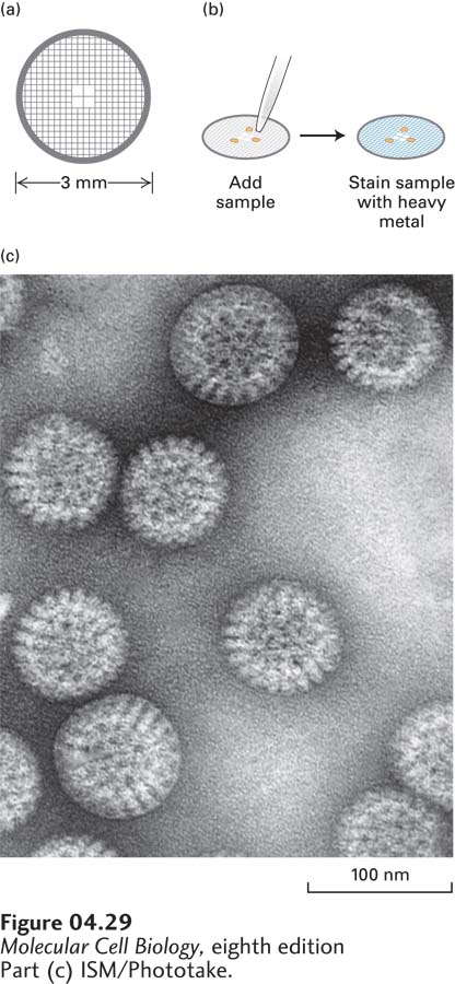
It is common in biology to explore the detailed shapes of single macromolecules, such as proteins or nucleic acids, or of structures, such as viruses and the filaments that make up the cytoskeleton. It is relatively easy to view these objects in the transmission electron microscope, provided they are stained with a heavy metal that scatters the incident electrons. To prepare a sample, it is first absorbed to a 3-mm electron microscope grid (Figure 4-29a), which is coated with a thin film of plastic and carbon. The sample is then bathed in a solution of a heavy metal, such as uranyl acetate, and excess solution is removed (Figure 4-29b). As a result of this procedure, the uranyl acetate coats the grid, but is excluded from the regions where the sample has adhered. When we view the sample in the TEM, we see where the stain has been excluded, so the sample is said to be negatively stained. Because the stain can precisely reveal the topology of the sample, a high-resolution image can be obtained (Figure 4-29c).
[Part (c) ISM/Phototake.]
FIGURE 4-29Transmission electron microscopy of negatively stained samples reveals fine features. (a) Samples for transmission electron microscopy (TEM) are usually mounted on a small copper or gold grid. The grid is usually covered with a very thin film of plastic and carbon to which a sample can adhere. (b) The specimen is then incubated in a heavy metal, such as uranyl acetate, and excess stain is removed. (c) The sample excludes the stain, so when it is observed in the TEM, it is seen in negative outline. The example in (c) is a negative stain of rotaviruses.
[Part (c) ISM/Phototake.]
Page 158
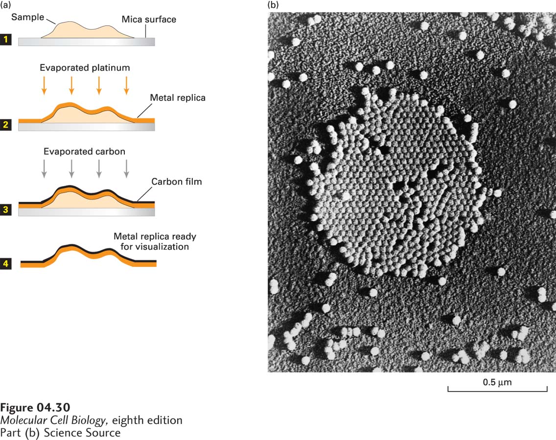
Samples can also be prepared by metal shadowing (Figure 4-30). In this technique, the sample is absorbed to a small piece of mica, then coated with a thin film of platinum by evaporation of the metal, then dissolved with acid or bleach, leaving the platinum coating (known as a replica). The platinum coating can be generated from a fixed angle or at a low angle as the sample is rotated, in which case it is called low-angle rotary shadowing. When the replica is transferred to a grid and examined in the TEM, it provides information about the three-dimensional topology of the sample.
[Part (b) Science Source]
FIGURE 4-30Metal shadowing makes surface details on very small objects visible by transmission electron microscopy. (a) The sample is spread on a mica surface and then dried in a vacuum evaporator (step 1). The sample grid is coated with a thin film of a heavy metal, such as platinum or gold, evaporated from an electrically heated metal filament (step 2). To stabilize the replica, the specimen is then coated with a carbon film evaporated from an overhead electrode (step 3). The biological material is then dissolved by acid and bleach (step 4), and the remaining metal replica is viewed in a TEM. In electron micrographs of such preparations, the carbon-coated areas appear light—the reverse of micrographs of simple metal-stained preparations, in which the areas of heaviest metal staining appear the darkest. (b) A platinum-shadowed replica of poliovirus particles.