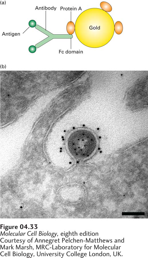
Immunoelectron Microscopy Localizes Proteins at the Ultrastructural Level
Just as immunofluorescence microscopy is used for localizing proteins at the light-microscope level, methods have been developed to use antibodies to localize proteins in thin sections at the electron microscope level. However, the harsh procedures used to prepare traditional thin sections—chemical fixation and embedding in plastic—can denature or modify the antigens so that they are no longer recognized by specific antibodies. Gentler methods, such as light fixation, sectioning of material after freezing at the temperature of liquid nitrogen, and finally incubation with antibody at room temperature, have been developed. To make the antibody visible in the electron microscope, it must be attached to an electron-dense marker. One way to do this is to use electron-dense gold particles coated with protein A, a bacterial protein that binds the Fc segment of all antibody molecules (Figure 4-33). Because the gold particles diffract incident electrons, they appear as dark spots.
[Courtesy of Annegret Pelchen-Matthews and Mark Marsh, MRC-Laboratory for Molecular Cell Biology, University College London, UK.]
FIGURE 4-33Gold particles coated with protein A are used to detect an antibody-bound protein by transmission electron microscopy. (a) First, antibodies are allowed to interact with their specific antigen in a section of fixed tissue. Then the section is treated with electron-dense gold particles coated with protein A from the bacterium S. aureus. Binding of the bound protein A to the Fc domains of the antibody molecules makes the location of the target protein visible in the electron microscope. (b) HIV particle budding from an infected HeLa cell. A cryosection of the specimen was prepared and first incubated with an antibody to capsid protein, then with protein A–coated 5-nm gold particles to localize the internal capsid protein. The unoccupied sites in the protein A were inactivated, and the specimen was incubated with antibody to the membrane-bound Env protein, followed by protein A–coated 10-nm gold particles. The distinct localization of the 5-nm gold labeling the capsid protein and the 10-nm gold labeling the Env protein can be seen. Scale bar is 100 nm.
[Courtesy of Annegret Pelchen-Matthews and Mark Marsh, MRC-Laboratory for Molecular Cell Biology, University College London, UK.]