The Folded Structure of tRNA Promotes Its Decoding Functions
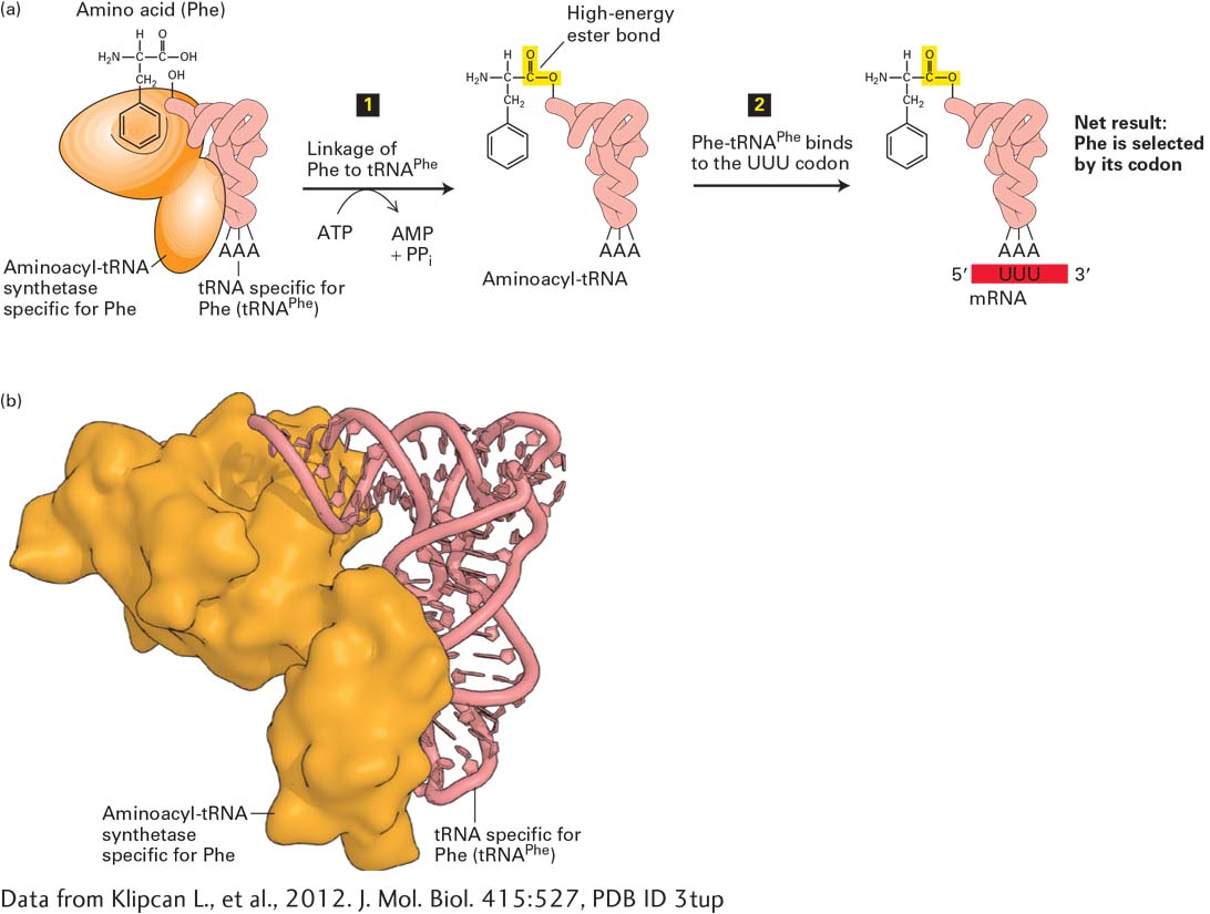
Translation, or decoding, of the four-nucleotide language of DNA and mRNA into the twenty–amino acid language of proteins requires both tRNAs and enzymes called aminoacyl-tRNA synthetases. To participate in protein synthesis, a tRNA molecule must become chemically linked to a particular amino acid via a high-energy bond, forming an aminoacyl-tRNA (Figure 5-19). The anticodon in the tRNA then base-pairs with a codon in mRNA so that the activated amino acid can be added to the growing polypeptide chain (see Figure 5-17).
[Data from Klipcan L., et al., 2012. J. Mol. Biol. 415:527, PDB ID 3tup.]
FIGURE 5-19Translating nucleic acid sequence into amino acid sequence. (a) The process for translating nucleic acid sequences in mRNA into amino acid sequences in proteins involves two steps. Step 1: An aminoacyl-tRNA synthetase first couples a specific amino acid, via a high-energy ester bond (yellow), to either the 2′ or 3′ hydroxyl of the terminal adenosine in the corresponding tRNA. Step 2: A three-base sequence in the tRNA (the anticodon) then base-pairs with a codon in the mRNA specifying the attached amino acid. If an error occurs in either step, the wrong amino acid may be incorporated into a polypeptide chain. Phe = phenylalanine. (Note that this is a simplified diagram of tRNAPhe ; its actual structure is shown in Figure 5-20b.) (b) Molecular model of the human mitochondrial aminoacyl-tRNA synthetase for Phe in complex with tRNAPhe.
[Data from Klipcan L., et al., 2012. J. Mol. Biol. 415:527, PDB ID 3tup.]
Some 30–40 different tRNAs have been identified in bacterial cells and as many as 50–100 in animal and plant cells. Thus the number of tRNAs in most cells is more than the number of amino acids used in protein synthesis (20) and also differs from the number of amino acid codons in the genetic code (61). Consequently, many amino acids have more than one tRNA to which they can attach (explaining how there can be more tRNAs than amino acids); in addition, many tRNAs can pair with more than one codon (explaining how there can be more codons than tRNAs).
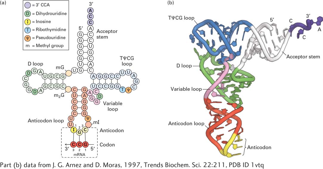
The function of tRNA molecules, which are 70–80 nucleotides long, depends on their precise three-dimensional structures. In solution, all tRNA molecules fold into a similar stem-loop arrangement that resembles a cloverleaf when drawn in two dimensions (Figure 5-20a). The four stems are short double helices stabilized by Watson-Crick base pairing; three of the four stems have loops containing seven or eight bases at their ends, while the remaining, unlooped stem contains the free 3′ and 5′ ends of the chain. The three nucleotides composing the anticodon are located at the center of the middle loop, in an accessible position that facilitates codon-anticodon base pairing. In all tRNAs, the 3′ end of the unlooped acceptor stem, to which a specific amino acid is attached, has the sequence CCA, which in most cases is added after synthesis and processing of the tRNA are complete. Several bases in most tRNAs are also modified after transcription, creating nonstandard nucleotides such as inosine, dihydrouridine, and pseudouridine. As we will see shortly, some of these modified bases are known to play an important role in protein synthesis. Viewed in three dimensions, the folded tRNA molecule has an L shape, with the anticodon loop and acceptor stem forming the ends of the two arms (Figure 5-20b).
Page 186
[Part (b) data from J. G. Arnez and D. Moras, 1997, Trends Biochem. Sci.22:211, PDB ID 1vtq.]
FIGURE 5-20Structure of tRNAs. (a) Although the exact nucleotide sequence varies among tRNAs, they all fold into four base-paired stems and three loops. The CCA sequence at the 3′ end is also found in all tRNAs. Attachment of an amino acid to the 3′ A yields an aminoacyl-tRNA. Some of the A, C, G, and U residues are modified post-transcriptionally in most tRNAs (see key). Dihydrouridine (D) is nearly always present in the D loop; likewise, ribothymidine (T) and pseudouridine (ψ) are almost always present in the TΨCG loop. Yeast alanine tRNA, represented here, also contains other modified bases. The triplet at the tip of the anticodon loop base-pairs with the corresponding codon in mRNA. See R. W. Holly et al., 1965, Science147:1462. (b) Three-dimensional model of the generalized backbone of all tRNAs. Note the L shape of the molecule.
[Part (b) data from J. G. Arnez and D. Moras, 1997, Trends Biochem. Sci.22:211, PDB ID 1vtq.]