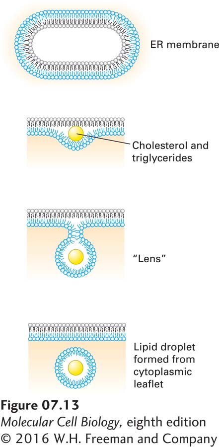
Lipid droplets are vesicular structures, composed of triglycerides and cholesterol esters, that originate from the ER and serve a lipid-storage function. When a cell’s supply of lipids exceeds the immediate need for membrane construction, excess lipids are relegated to these lipid droplets, readily visualized in live cells by staining with a lipophilic dye such as Congo red. Feeding cells with oleic acid, a type of fatty acid, enhances lipid droplet formation. Lipid droplets are not only storage compartments for triglycerides and cholesterol esters, but may also serve as platforms for storage of proteins targeted for degradation. The biogenesis of lipid droplets starts with delamination of the lipid bilayer of the ER through insertion of triglycerides and cholesterol esters (Figure 7-13). The lipid “lens” continues to grow by insertion of more lipid, until finally a lipid droplet is hatched by scission from the ER. The resulting cytoplasmic droplet is thereby surrounded by a phospholipid monolayer. The details of lipid droplet biogenesis, as well as its functions, remain to be defined more clearly.
FIGURE 7-13Lipid droplets form by budding and scission from the ER membrane. Lipid droplet formation begins with the accumulation of cholesterol esters and triglycerides (yellow) within the hydrophobic core of the lipid bilayer. The resulting delamination of the two lipid monolayers causes a “lens” to form, the further growth of which creates a spherical droplet that is then released by scission at the neck. The newly formed droplet is surrounded by a lipid monolayer, derived from the cytosolic leaflet of the ER membrane.