Histones, the most abundant proteins in chromatin, constitute a family of small, basic proteins. The five major types of histone proteins—termed H1, H2A, H2B, H3, and H4—are rich in positively charged basic amino acids, which interact with the negatively charged phosphate groups in DNA.
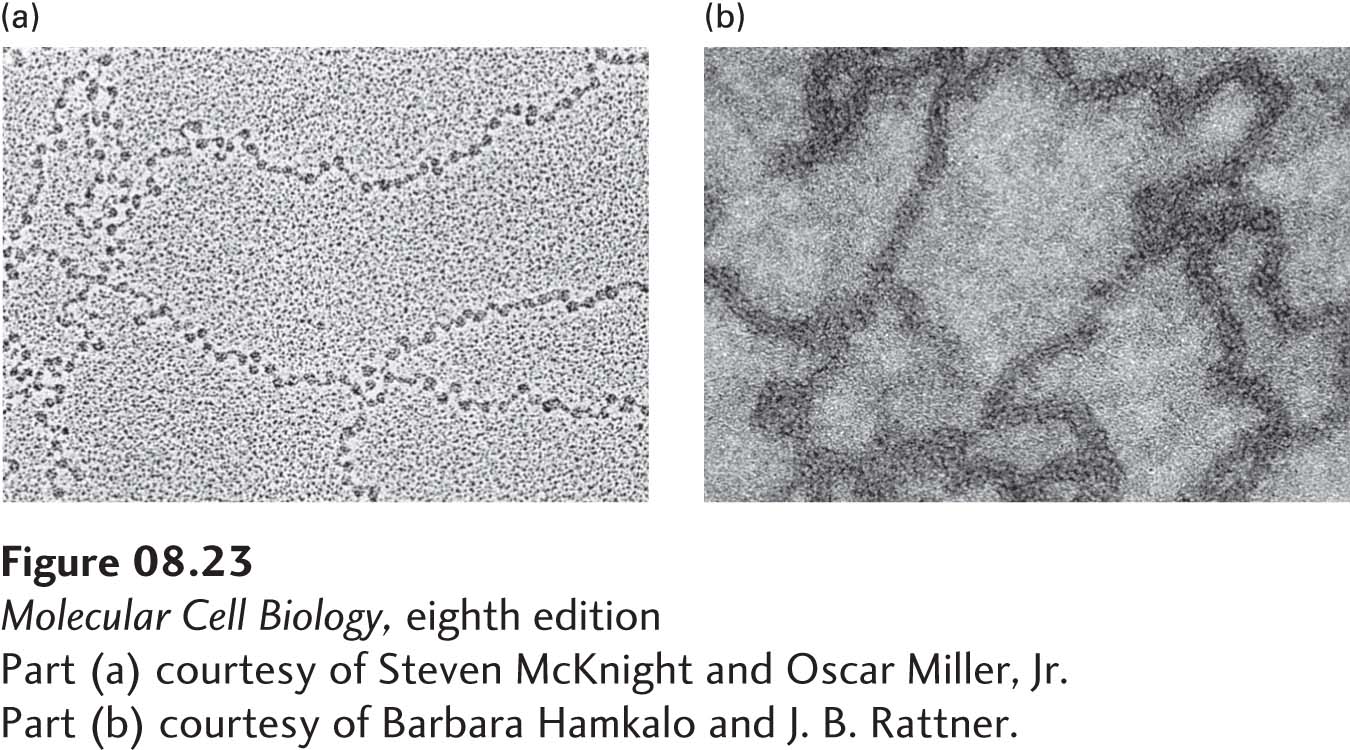
When chromatin is extracted from nuclei and examined in the electron microscope, its appearance depends on the salt concentration to which it is exposed. At low salt concentrations and in the absence of divalent cations such as Mg2+, isolated chromatin resembles beads on a string (Figure 8-23a). In this extended form, the “string” is composed of free DNA, called “linker” DNA, connecting beadlike structures termed nucleosomes. Composed of DNA and histones, nucleosomes are about 10 nm in diameter and are the primary structural units of chromatin. If chromatin is isolated at a physiological salt concentration, it assumes a more condensed fiberlike form that is about 30 nm in diameter (Figure 8-23b).
[Part (a) courtesy of Steven McKnight and Oscar Miller, Jr. Part (b) courtesy of Barbara Hamkalo and J. B. Rattner.]
EXPERIMENTAL FIGURE 8-23The extended and condensed forms of extracted chromatin have very different appearances in electron micrographs. (a) Chromatin isolated in low-ionic-strength buffer has an extended “beads-on-a-string” appearance. The “beads” are nucleosomes (10 nm in diameter) and the “string” is connecting (linker) DNA. (b) Chromatin isolated in buffer with a physiological ionic strength (0.15 M KCl) appears as a condensed fiber 30 nm in diameter.
[Part (a) courtesy of Steven McKnight and Oscar Miller, Jr. Part (b) courtesy of Barbara Hamkalo and J. B. Rattner.]
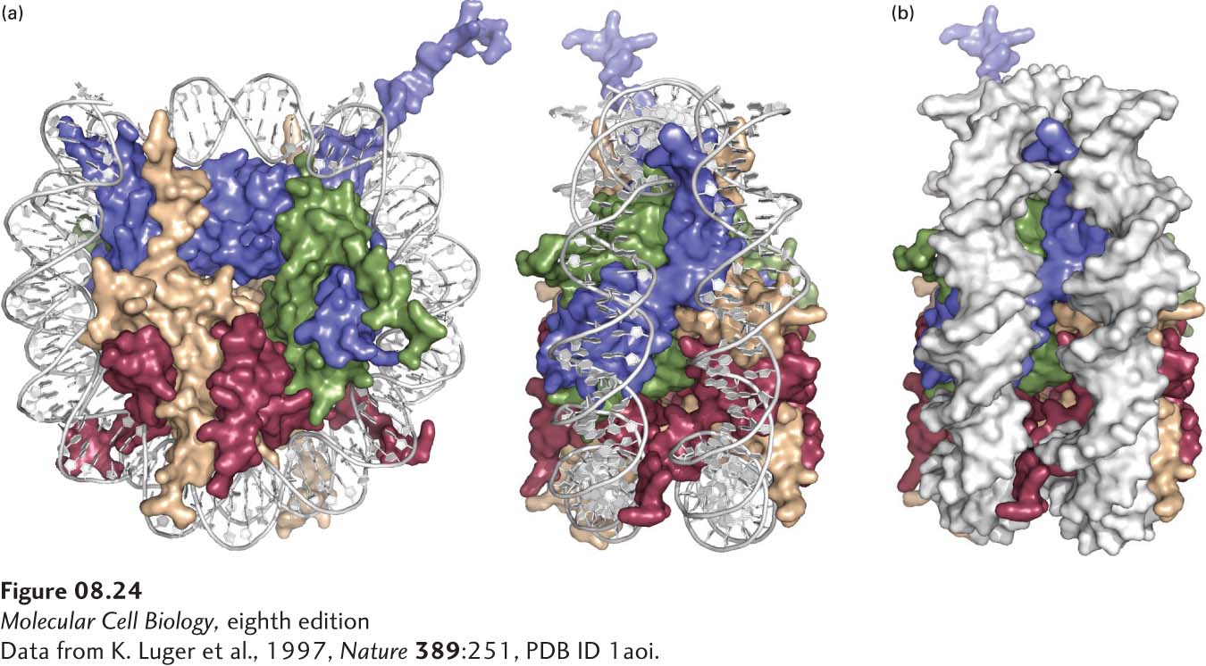
Structure of Nucleosomes The DNA component of nucleosomes is much less susceptible to nuclease digestion than is the linker DNA between them. If nuclease treatment is carefully controlled, all the linker DNA can be digested, releasing individual nucleosomes with their DNA component. A nucleosome consists of a protein core with DNA wound around its surface like thread around a spool. The core is an octamer containing two copies each of histones H2A, H2B, H3, and H4. X-ray crystallography has shown that the octameric histone core is a roughly disk-shaped structure made of interlocking histone subunits (Figure 8-24). Histones H3 and H4 fold into a tetramer in the absence of histones H2A and H2B. Two heterodimers of H2A and H2B then associate with the H3-H4 tetramer. Positive charges on the surface of the histone octamer in the region interacting with DNA hold the negatively charged DNA against the surface of the histone octamer at the center of the nucleosome. Nucleosomes from all eukaryotes contain about 147 bp of DNA wrapped one and two-thirds turns around the globular histone core. The length of the linker DNA is more variable among species, and even between different cells of the same organism, ranging from about 10 to 90 base pairs. During cell replication, DNA is assembled into nucleosomes shortly after the replication fork passes (see Figure 5-32). This process depends on specific chaperone molecules that bind to histones and assemble them, together with newly replicated DNA, into nucleosomes.
[Data from K. Luger et al., 1997, Nature389:251, PDB ID 1aoi.]
FIGURE 8-24Structure of the nucleosome based on x-ray crystallography. (a) Nucleosome with space-filling model of the histones. The sugar-phosphate backbones of the DNA strands are represented as white tubes to allow better visualization of the histones. Nucleosome shown from the top (left) and from the side (right, rotated clockwise 90°). H2A subunits are yellow; H2Bs are red; H3s are blue; H4s are green. The N-terminal tails of the eight histones and the H2A and H2B C-terminal tails, involved in condensation of the chromatin, are not visible because they are disordered in the crystal. One H2A, H2B heterodimer projects out of the page on the lower right of the side view, while the other H2A, H2B heterodimer projects into the page, on the lower left of the side view. Only one H2A, H2B heterodimer is visible in the top view. The other H2A, H2B dimer is not visible in this view because it is behind the H3, H4 tetramer, on the lower right. (See also the ribbon diagram of the histone polypeptide chains in Figure 8-26, where only one H2A, H2B heterodimer is clearly visible on the lower left of the top view of the nucleosome.) (b) Space-filling model of histones and DNA (white) viewed from the side of the nucleosome. See also http://lugerlab.org for a rotating movie of the nucleosome core.
[Data from K. Luger et al., 1997, Nature389:251, PDB ID 1aoi.]
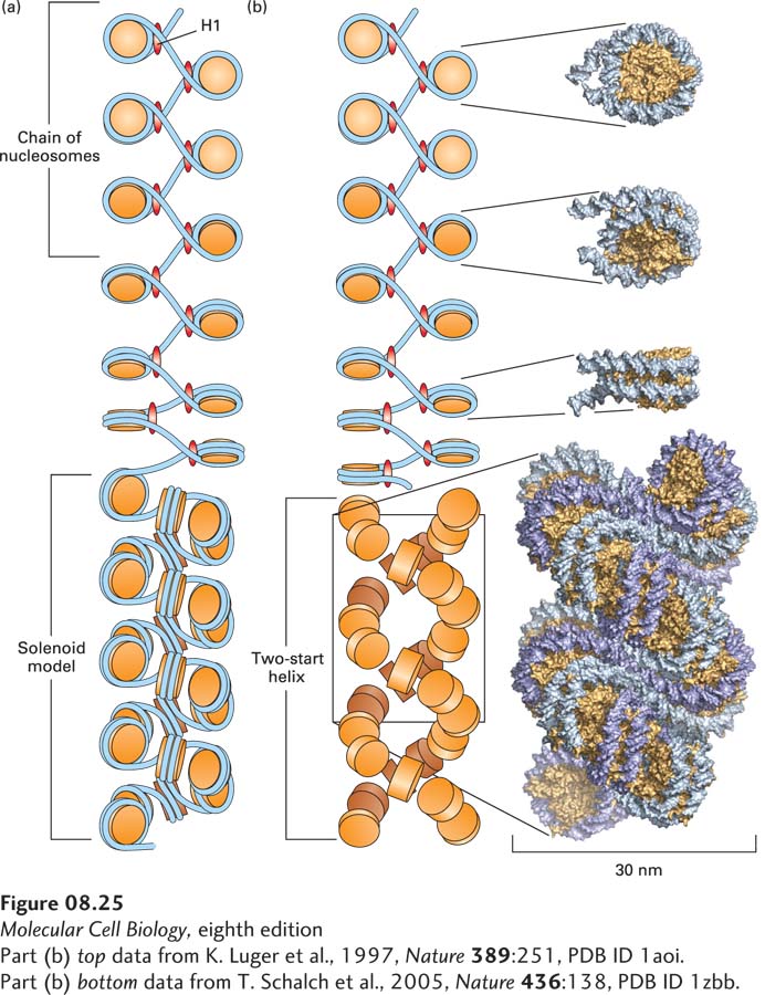
Structure of the 30-nm Fiber When extracted from cells in isotonic buffers (i.e., buffers with the same salt concentration found in cells, about 0.15 M KCl, 0.004 M MgCl2), most chromatin appears as fibers about 30 nm in diameter (see Figure 8-23b). Despite many years of study, the arrangement of nucleosomes in the 30-nm chromatin fiber remains controversial. In one widely accepted model with considerable experimental support, nucleosomes are arranged in a helical conformation forming a solenoid with six or more nucleosomes per turn of the helix (Figure 8-25a). In a second strongly supported model, the two-start helix, nucleosomes alternately stack like coins into two “strands,” and those strands wind into a left-handed double helix (Figure 8-25b). The 30-nm fibers also include H1, the fifth major histone. H1 is bound to the DNA as it enters and exits the nucleosome core, but its structure in the 30-nm fiber is not known at atomic resolution.
[Part (b) top data from K. Luger et al., 1997, Nature389:251, PDB ID 1aoi. Part (b) bottom data from T. Schalch et al., 2005, Nature436:138, PDB ID 1zbb.]
FIGURE 8-25Models of the structure of the 30-nm chromatin fiber. (a) In the solenoid model, nucleosomes are arranged in a left-handed helix with six or more nucleosomes per turn. See M. Kruithof et al., 2009, Nature Struc. Mol. Biol.16:534. (b) In the two-start helix model, a “zigzag ribbon” of nucleosomes (top) folds into a two-start helix (bottom). For simplicity, DNA is not represented in the two-start helix. See C. L. F. Woodcock et al., 1984, J. Cell Biol.99:42.
[Part (b) top data from K. Luger et al., 1997, Nature389:251, PDB ID 1aoi. Part (b) bottom data from T. Schalch et al., 2005, Nature436:138, PDB ID 1zbb.]
Page 329
The chromatin in chromosomal regions that are not being transcribed or replicated exists predominantly in the condensed, 30-nm fiber form and in higher-order folded structures whose detailed conformation is not currently understood. The regions of chromatin actively being transcribed and replicated are thought to assume the extended beads-on-a-string form.
Conservation of Chromatin Structure The general structure of chromatin is remarkably similar in the cells of all eukaryotes, including fungi, plants, and animals, indicating that the structure of chromatin was optimized early in the evolution of eukaryotic cells. The amino acid sequences for the four core histones (H2A, H2B, H3, and H4) are highly conserved between distantly related species. For example, the sequences of histone H3 from sea urchin tissue and calf thymus differ by only a single amino acid, and H3 from the garden pea and calf thymus differ by only four amino acids. Apparently, significant deviations from the histone amino acid sequences were selected against strongly during evolution. The amino acid sequence of H1, however, varies more from organism to organism than do the sequences of the other major histones. The similarity in sequence among histones from all eukaryotes indicates that they fold into very similar three-dimensional conformations, which were optimized for histone function early in evolution in a common ancestor of all modern eukaryotes.
Minor histone variants encoded by genes that differ from the highly conserved major types also exist, particularly in vertebrates. For example, a special form of H2A, designated H2AX, is incorporated into nucleosomes in place of H2A in a small fraction of nucleosomes in all regions of chromatin. At sites of double-stranded breaks in chromosomal DNA, H2AX becomes phosphorylated and participates in the chromosome-repair process, probably by functioning as a binding site for repair proteins. In the nucleosomes at centromeres, H3 is replaced by another variant histone called CENP-A, which participates in the binding of spindle microtubules during mitosis. A variant of histone H3, known as H3.3, replaces the major histone H3 in transcribed regions of DNA, probably when the histone octamer must be moved out of the way by histone chaperones as RNA polymerase transcribes the DNA in chromatin. Most minor histone variants differ only slightly in sequence from the major histones. These slight changes in histone sequence may influence the stability of the nucleosome as well as its tendency to fold into the 30-nm fiber and other higher-order structures.