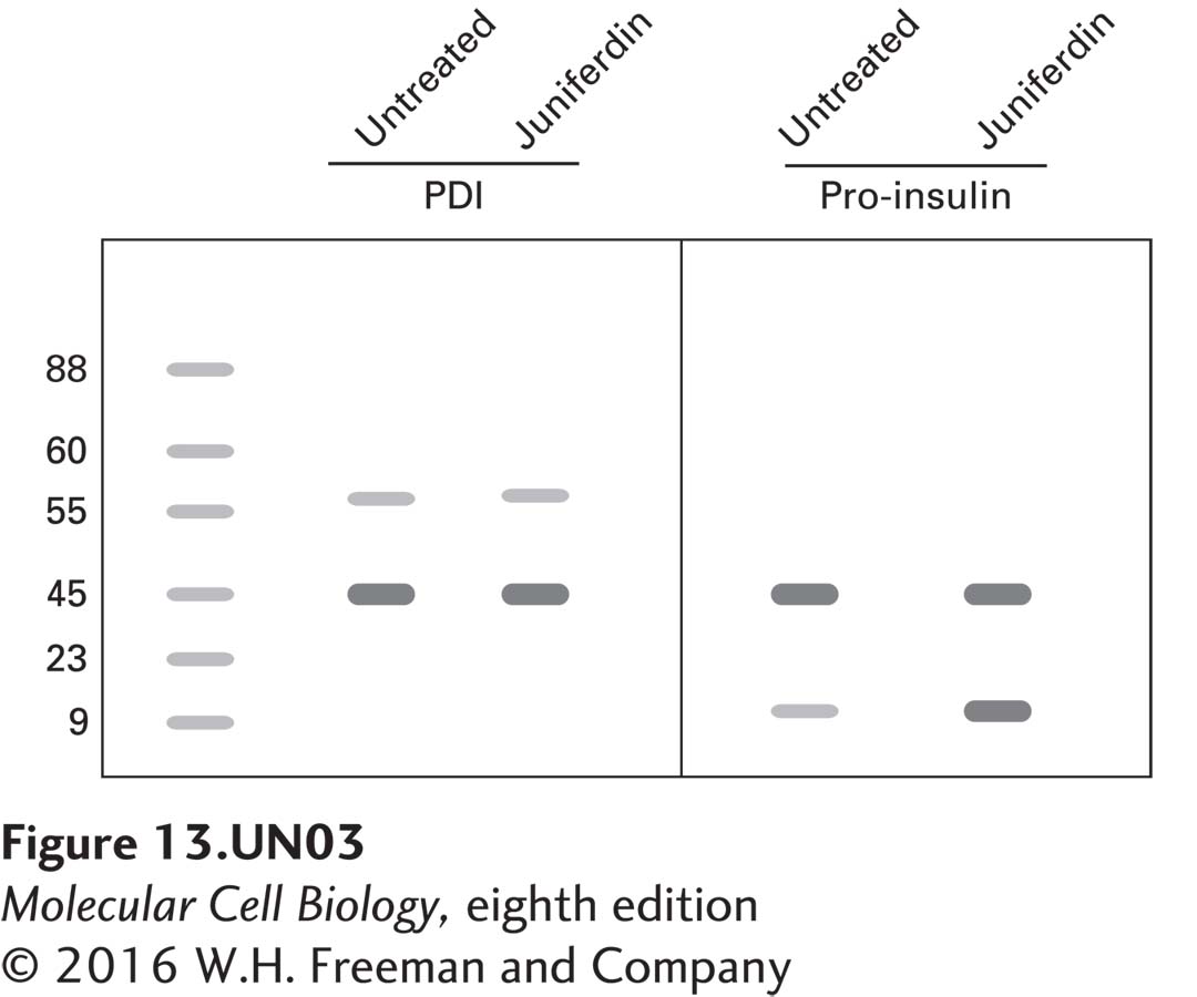
Recently, researchers discovered that treating mammalian cells with juniferdin, a plant-derived compound, affects protein secretion, and they have reported that the target of this drug is protein disulfide isomerase (PDI). In the following experiment, cultured pancreatic β-cells were treated with juniferdin, and protein lysates were isolated and compared with lysates from untreated cells using immunoblot analysis. Probing of blots with antibodies against PDI (57 kDa), actin (43 kDa), and proinsulin (9.8 kDa) shows the following:
a. Given that approximately the same amount of protein was loaded in each lane, as evidenced by the actin signals, how do you explain the fact that the PDI levels also appear about the same, while most of the proinsulin remains accumulated in the juniferdin-treated cells?
The fact that pro-insulin accumulates indicates that it has not folded properly because of the inability of PDI to arrange the correct disulfide bonding needed for processing to the Golgi and its eventual secretion as insulin. That PDI is present in equal amounts indicates that juniferdin has no effect on PDI synthesis; rather, it works to block the activity of PDI as an enzyme, which explains the pro-insulin accumulation.
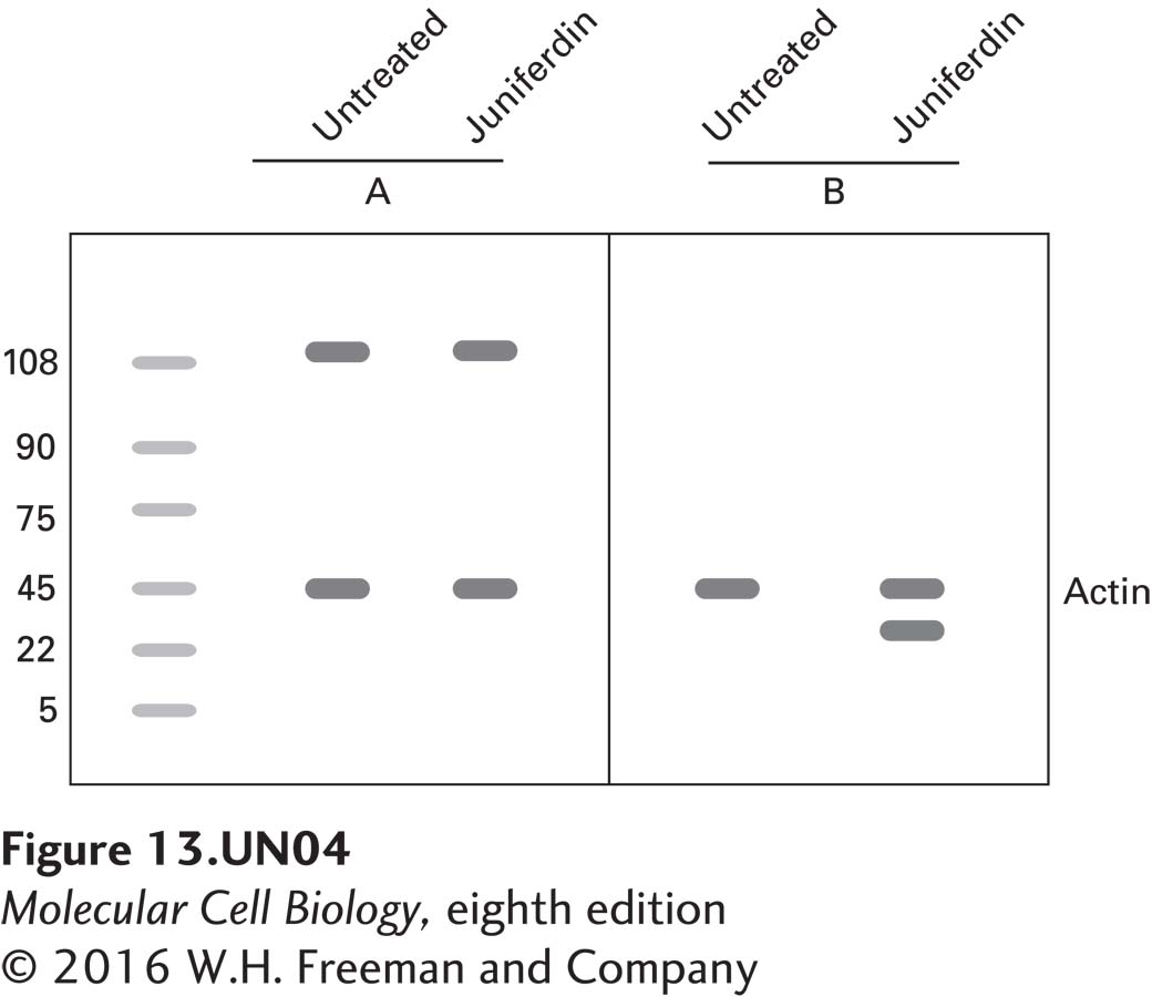
b. To confirm these results, protein lysates of juniferdintreated and untreated cells were separated by SDS-PAGE, blotted to membranes, and then probed with antibodies against Ire1 and Hac1.
Label each blot with the antibody that was used for the analysis. How do you explain the increase in the intensity of the signal seen in the juniferdin-treated cells in blot B?
A is Ire1, which would not change due to treatment, and because of the pro-insulin pile-up, it would lose its association with Bip, which is now bound to the improperly folded pro-insulin. Ire1 not bound to Bip dimerizes into an active endonuclease, which leads to the processing of the Hac1 mRNA and its translation into protein. Hence, increased the levels of Hac1 on blot B.
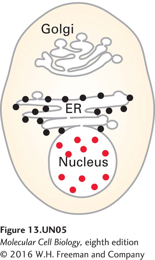
c. An immunocytochemistry and fluorescence microscopy analysis was undertaken with the antibody used in blot B and a secondary antibody labeled with rhodamine (red). Since juniferdin specifically affects PDI, a resident rough ER (RER) protein, how do you explain the localization of the signal in the nucleus?
The increase in Hac1 leads to its translocation to the nucleus where it serves in the transcription of genes encoding proteins that will assist in folding and the progression of proteins through to the Golgi and their eventual secretion.