
Chapter 14. Analyzing I-Cell Disease Symptoms
Introduction
Analyze the Data 14-3: Analyzing I-Cell Disease Symptoms
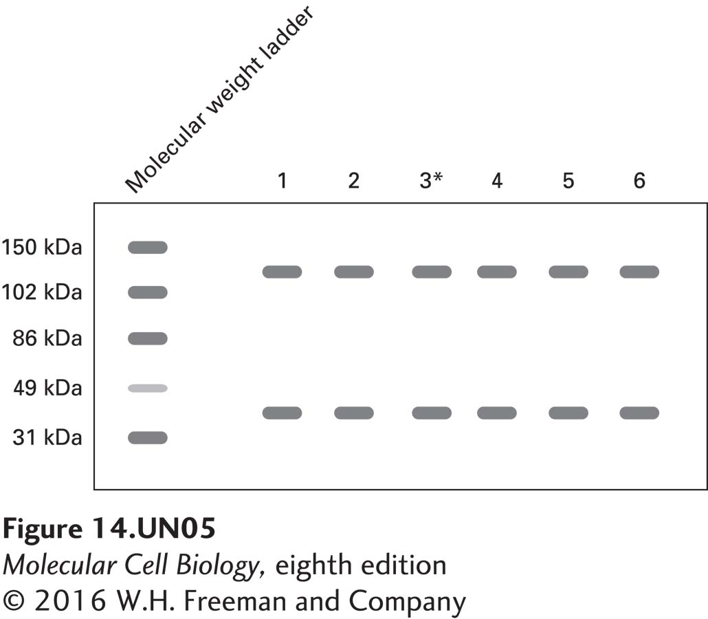
A child appears to be suffering from I-cell disease, but when a sample of his proteins (lane 3* below), isolated from skin fibroblasts, is compared with protein samples from fibroblasts of his healthy parents (lanes 1 and 2) and siblings (lanes 4–6) using Western blot analysis and antibodies against N-acetylglucosamine phosphotransferase (~145 kDa) and actin (loading control, ~43 kDa), the following pattern is seen:
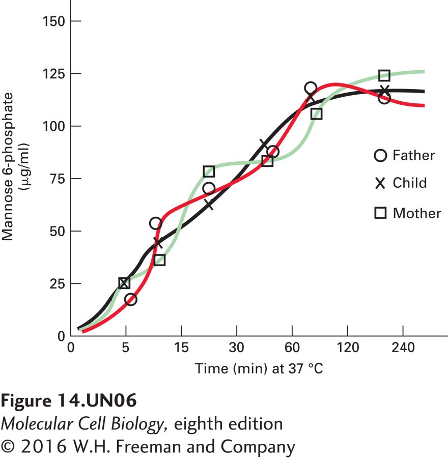
In a second set of experiments, N-acetylglucosamine phosphotransferase was isolated from cells from the afflicted child and from his healthy parents and used in an assay with 32P to measure enzyme activity and the production of mannose 6-phosphate. The assay yielded the following results:
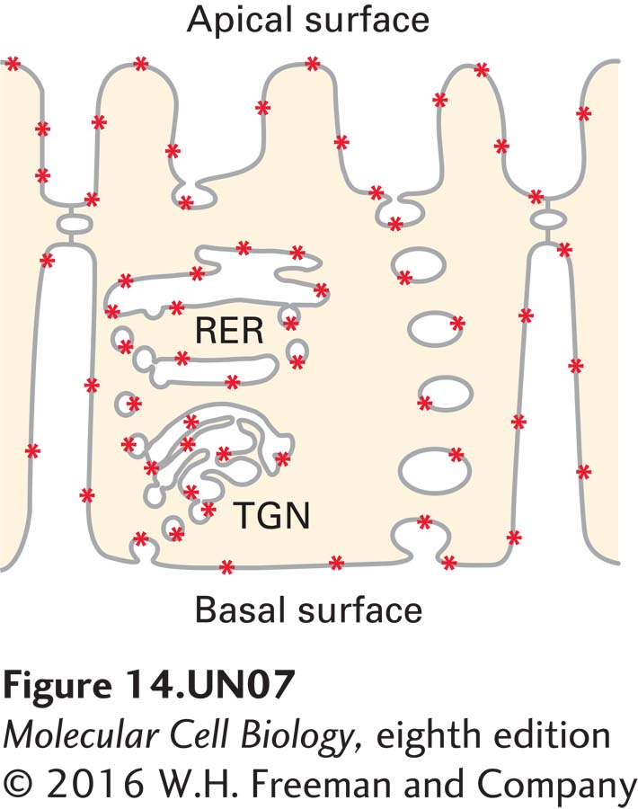
a. Using fibroblasts cultured from the child, design an experiment using the N-acetylglucosamine phosphotransferase antibody and fluorescence microscopy and describe the results that could explain why the child presents symptoms similar to those of I-cell disease.
The cell should show fluorescence in the RER and nowhere else. The reason is that a) the protein is there as evident by Western blot and b) it is biologically active as evident by the assay above. The only explanation for the symptoms, then, is that the protein is not getting to the right place (i.e., the cis-Golgi), which is why there are no lysosomal enzymes modified with M6P and hence the I-cell symptoms.
Activity results are being submitted...