HOW DO WE KNOW?
What are the shapes of proteins?
BACKGROUND The three-dimensional shapes of proteins can be determined by X-ray crystallography. One of the pioneers in this field was Dorothy Crowfoot Hodgkin, who used this technique to define the structures of cholesterol, vitamin B12, penicillin, and insulin. She was awarded the Nobel Prize in Chemistry in 1964 for her early work. Max Perutz and John Kendrew shared the Nobel Prize in Chemistry in 1962 for defining the structures of myoglobin and hemoglobin using this method.
METHOD X-ray crystallography can be used to determine the shape of proteins, as well as other types of molecules. The first step, which can be challenging, is to make a crystal of the protein molecules. A crystal is a solid structure in which the atoms of a protein (or any other molecule) are in an ordered and repeating pattern in three dimensions. Then X-rays are aimed at the crystal while it is rotated. Some X-rays pass through the crystal, while others are scattered in different directions when they hit atoms of the proteins. A film or other detector records the pattern as a series of spots, which is known as a diffraction pattern. The locations and intensities of these spots can be used to infer the position and arrangement of the atoms in the molecule.
RESULTS The X-ray diffraction pattern for hemoglobin looks like this:
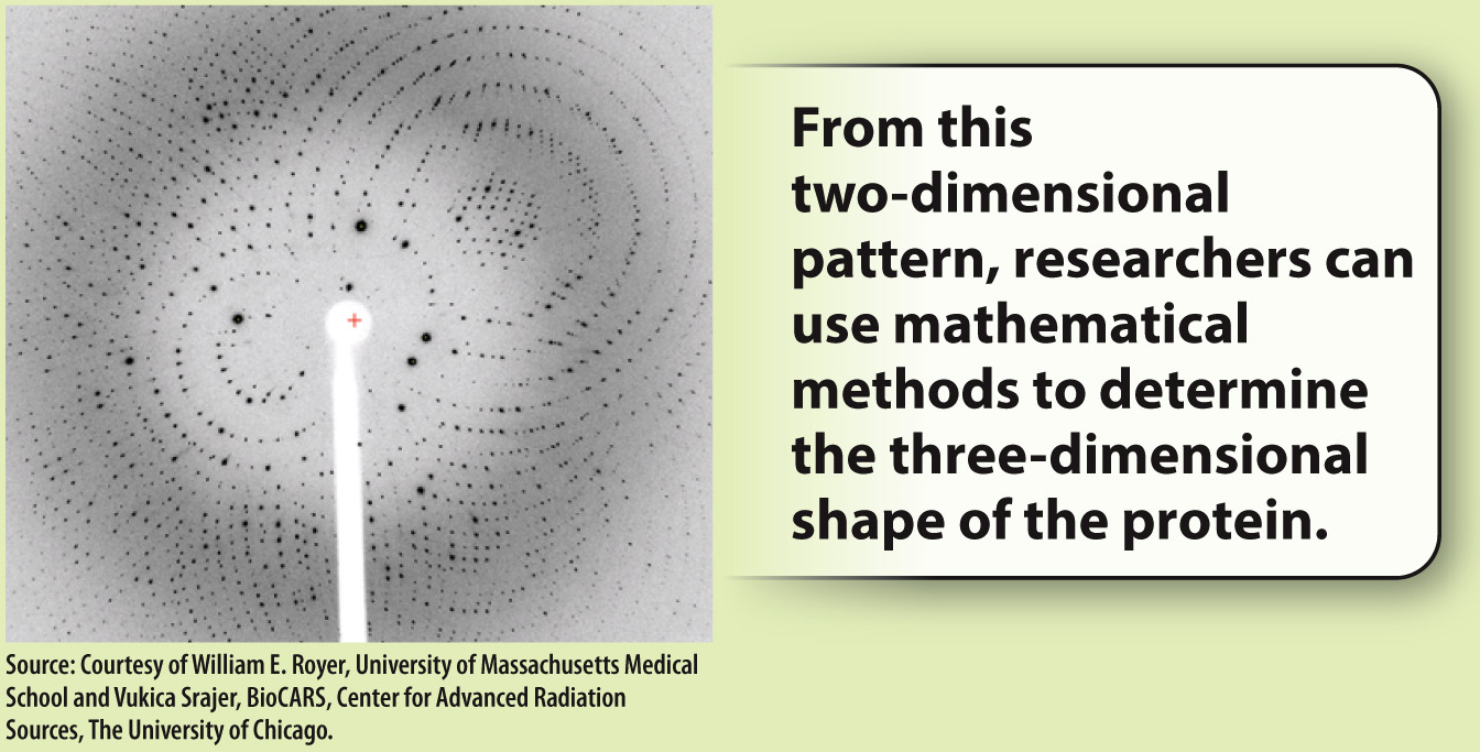
FIG. 4.5
FOLLOW-UP WORK Linus Pauling and Robert Corey used X-ray crystallography to determine two types of secondary structures commonly found in proteins—the α helix and the β sheet. Today, this technique is a common method for determining the shape of proteins.
SOURCES Crowfoot, D. 1935. “X-Ray Single Crystal Photographs of Insulin.” Nature 135:591–592; Kendrew, J. C., et al. 1958. “A Three-Dimensional Model of the Myoglobin Molecule Obtained by X-Ray Analysis.” Nature 181:662–666.