Motor proteins associate with microtubules and microfilaments to cause movement.
A motor is a device that imparts motion. We saw that microtubules and microfilaments have some capacity to lengthen and shorten by polymerization and depolymerization. However, when joined by small accessory proteins called motor proteins, microtubules and microfilaments are capable of causing amazing movements.
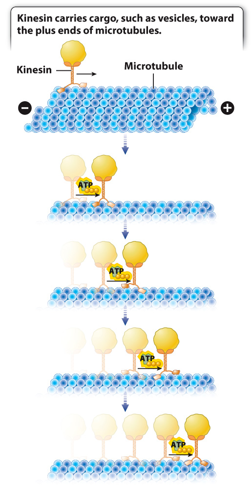
For example, microtubules function as tracks for transport within the cell. Two motor proteins that associate with these microtubule tracks are kinesin and dynein. Kinesin transports cargo toward the plus end of microtubules, located at the periphery of the cell (Fig. 10.7). By contrast, dynein carries its load away from the plasma membrane toward the minus end, located at the centrosome in the interior of the cell. Movement along microtubules by kinesin and dynein is driven by conformational changes in the motor proteins and is powered by energy harvested from ATP.
Let’s look at an especially striking example of this system at work in the specialized skin cells called melanophores present in some vertebrates. Melanophores are similar to the melanocytes in our own skin that produce the pigment melanin. However, rather than hand off their melanin to other cells as in humans, melanophores keep their pigment granules and move them around the cell in response to hormones or neuronal signals. This redistribution of melanin within the cell allows animals such as fish or amphibians to change color. For example, at night the melanin granules in the skin of a zebrafish embryo are dispersed throughout the melanophores, making it darkly colored. As morning comes and the day brightens, the pigment granules aggregate at the center of the cell around the centrosome, causing the embryo’s color to lighten (Fig. 10.8).
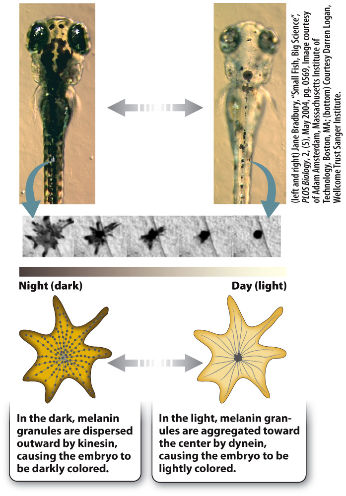
The melanin granules in the melanophores move back and forth along microtubules, transported by kinesin and dynein. Kinesin moves the granules out toward the plus end of the microtubule during dispersal, and dynein moves them back toward the minus end during aggregation. The daytime and nighttime camouflage provided by this color change helps prevent young, developing organisms from being spotted by hungry predators lurking below.
Quick Check 1 Would a defect in dynein or in kinesin cause a zebrafish embryo to remain darkly colored after daybreak?
Quick Check 1 Answer
A defect in dynein would cause the melanin granules to remain dispersed because dynein transports the granules back toward the minus end of the microtubule during granule aggregation.
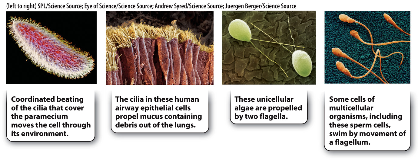
In addition to providing tracks for the transport of material within the cell, microtubules are found in cilia and flagella, fiberlike organelles that propel the movement of cells or substances surrounding the cell (Fig. 10.9). In these organelles, microtubules associate with the motor protein dynein, which causes movement. Many single-
Like microtubules, microfilaments also associate with motor proteins to produce movement. Actin microfilaments associate with myosin to transport various types of cellular cargo, such as vesicles, inside of cells. Furthermore, microfilaments are responsible for changes in the shape of many types of cell. One of the most dramatic examples of cell shape change is the shortening (contraction) of a muscle cell. Muscle contraction depends on the interaction of myosin with microfilaments, and is powered by ATP (Chapter 37).