Amoebozoans include slime molds that produce multicellular structures.
Previously in this chapter, we learned one thing about amoebas: they move and feed by extending cytoplasmic fingers called pseudopodia. As its name implies, the superkingdom Amoebozoa (Fig. 27.11a) is a group of eukaryotes with amoeba-like cells (illustrated by Amoeba proteus in Fig. 27.11b). More than 1000 amoebozoan species have been described. In some amoebozoan species, large numbers of cells aggregate to form multicellular structures as part of their life cycle.
Page 565
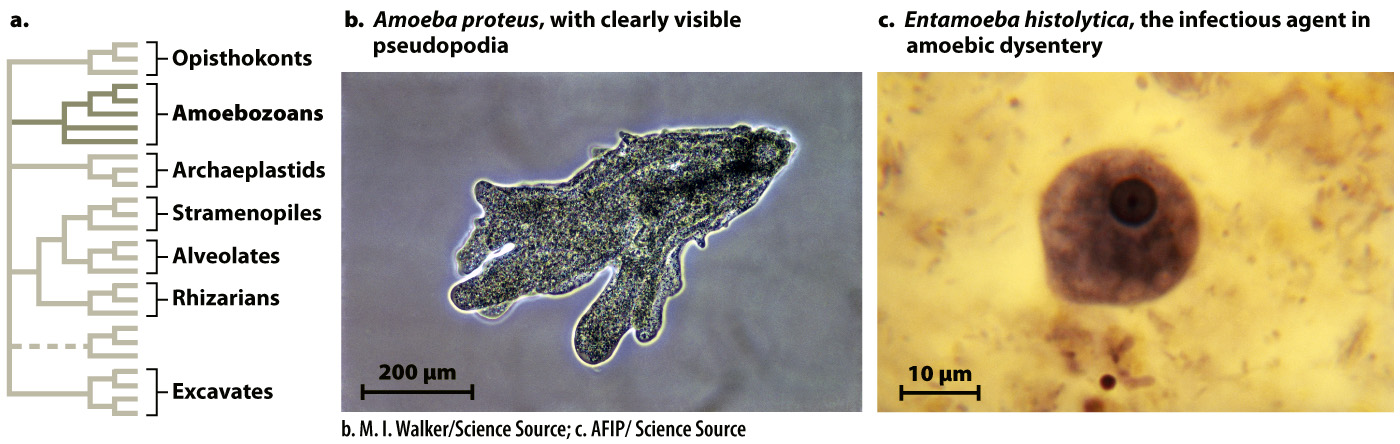
FIG. 27.11 Amoebozoans. This group includes protists with an amoeboid stage in their life cycle.
Photo sources: b. M. I. Walker/Science Source; c. AFIP/Science Source.
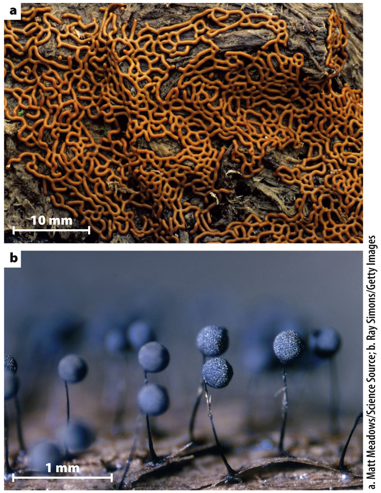
FIG. 27.12 Plasmodial slime molds. (a) Plasmodia, such as this pretzel slime mold, are coenocytic structures containing many nuclei. (b) Plasmodia generate sporangia, stalked structures that produce spores for dispersal.
Although there are amoeba-like cells that are not members of the Amoebozoa superkingdom, all members of this group have cells similar to that of Amoeba, at least at some stage of their life cycle. Amoebozoans play an important role in soils as predators on other microorganisms. Some amoebozoans cause disease in humans. For example, Entamoeba histolytica (Fig. 27.11c), an anaerobic protist that causes amoebic dysentery, is responsible for 50,000–100,000 deaths every year.
Beyond considerations of human health, the amoebozoans of greatest biological interest are the slime molds. In plasmodial slime molds (Fig. 27.12), haploid cells fuse to form zygotes that subsequently undergo repeated rounds of mitosis but not cell division to form colorful, often lacy structures visible to the naked eye (Fig. 27.12a). These structures, called plasmodia, are coenocytic, which means they contain many nuclei within one giant cell. The plasmodia can move and so seek out and feed on the bacteria and small fungi commonly found on bark or plant litter on the forest floor. Triggered by poorly understood environmental signals, plasmodia eventually differentiate to form stalked structures called sporangia that can be 1 to 2 mm high (Fig. 27.12b). Within the mature sporangium, cell walls form around the many nuclei, producing discrete cells. These cells undergo meiosis, generating haploid spores that disperse into the environment. Germination of these spores begins the life cycle anew.
Cellular slime molds (Fig. 27.13) are a second type of slime mold. These slime molds spend most of their life cycle as solitary amoeboid cells feeding on bacteria in the soil. Starvation, however, causes these cells to produce the chemical signal cyclic AMP (Chapter 9), which induces as many as 100,000 cells to aggregate into a large multicellular slug-like form (Fig. 27.13a). This “slug” can migrate by a coordinated movement of its cells governed by actin and myosin, the same proteins that control muscle movement in animals. Migrating “slugs” can forage for food, leaving behind a trail of mucilage as they move—that’s why they’re called slime molds. Like plasmodial slime molds, the “slugs” of cellular slime molds eventually differentiate to form stalk-borne sporangia that produce the spores responsible for dispersal of the organism and renewal of the life cycle (Fig. 27.13b).
Page 566
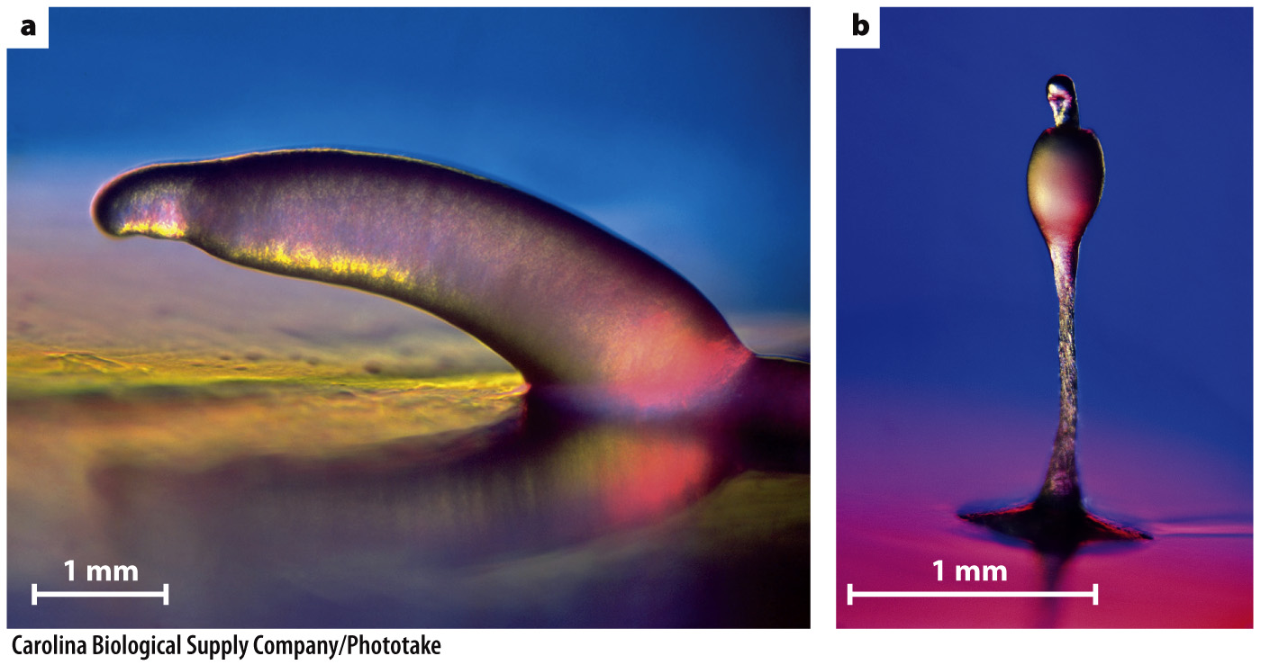
FIG. 27.13 Cellular slime molds. (a) Starvation causes amoeboid feeding cells of cellular slime molds to aggregate into a multicellular “slug” that can migrate along surfaces, leaving behind a trail of slime. (b) “Slugs” differentiate to form stalked sporangia that produce spores.
Slime molds are not true intermediates in the evolution of multicellularity—that is, they are not closely related to animals, fungi, or other groups in which complex multicellularity evolved. Nonetheless, the cellular slime mold Dictyostelium has been extensively studied by biologists as a model organism because it has much to teach us about fundamental eukaryotic processes of cell signaling and differentiation, cellular motility, and mitosis.