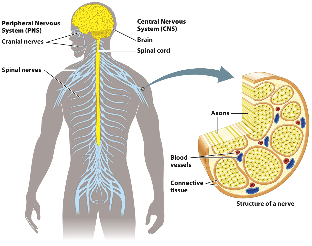
Nervous systems are organized into peripheral and central components.
As animals evolved the ability to sense and coordinate responses to increasingly complex stimuli, their nervous systems became organized into peripheral and central components (Fig. 35.16). Your eyes, receptors for the sense of touch, and other sensory organs are located on the surface of the body, where they can receive signals from the environment. In contrast, the brain, centrally located ganglia, and a main nerve cord are located in the interior.

Nerves form the lines of communication between these nervous system structures. In general, nerve cell bodies are grouped together in sensory organs, ganglia, and a main nerve cord that extends from the brain. Nerves are composed mainly of axons from many different nerve cells. For example, the optic nerve contains axons that travel from nerve cells in the eye to the brain.
Sensory and motor nerves make up the peripheral nervous system (PNS). These nerves communicate with the central nervous system (CNS), made up of the brain, a main nerve cord, or—
The peripheral nervous system also includes interneurons and ganglia that integrate and process information in local regions of the animal’s body. For example, vertebrates and invertebrates have ganglia that lie outside segments of their primary nerve cord that coordinate localized function within a region of their body, such as the muscles that control the limb of a running insect, the muscles that allow an earthworm to dig, or the secretory activity of a particular region of the digestive system of a fish or a mammal. However, the bulk of information processing, particularly in animals that exhibit a greater capacity for learning and memory, occurs in the CNS.
Let’s look more closely at the organization of the vertebrate nervous system. The central nervous system of vertebrates includes both the brain and spinal cord (Fig. 35.16). The spinal cord is a central tract of neurons that passes through the vertebrae to transmit information between the brain and the periphery of the body. The vertebrate spinal cord is divided into segments, each controlling body movement in a particular region along the animal’s length. Each spinal cord segment contains axons from peripheral sensory neurons, a set of interneurons, and a set of motor neuron cell bodies. These are distinct from, but often associated with, the ganglia mentioned above that lie outside the spinal cord.
In humans and other vertebrate animals, the peripheral nervous system is organized into left and right sets of cranial nerves located within the head and spinal nerves running from the spinal cord to the periphery. Most of the cranial nerves and all of the spinal nerves contain axons of both sensory and motor neurons. Cranial nerves link specialized sensory organs (eyes, ears, tongue) to the brain. Cranial nerves also control eye movement, facial expression, speech, and feeding. Some cranial nerves, such as the olfactory and optic nerves, contain only sensory axons. Spinal nerves exit from the spinal cord through openings located between adjacent vertebrae to thread through the trunk and limbs of an animal’s body. These nerves receive sensory information from receptors in nearby body regions along the length of the body and send motor signals from the spinal cord back to those regions (Fig. 35.16).