Simple reflex circuits provide rapid responses to stimuli.
An animal that has perceived a predator has an advantage if it can move quickly. Fast responses are made possible by simple reflex circuits that bypass the brain. These circuits connect sensory neurons (detecting the presence of the predator) directly with motor neurons (providing the quick movement necessary to escape the predator). Reflex circuits are common in both the somatic and autonomic components of the nervous system.
Simple spinal reflex circuits connect sensory and motor neurons directly in the spinal cord of vertebrates. The large Mauthner neurons of fish are an example of such a circuit. Sensory neurons respond to threatening visual, tactile, or vibratory cues from the environment and transmit signals through the spinal cord to the hindbrain of a fish, activating a large Mauthner neuron on the side opposite the stimulus. The Mauthner cell in turn rapidly activates motor neurons along the body, causing the fish to bend away from the threatening stimulus to initiate a rapid escape. At the same time, the Mauthner cell and motor nerves on the other side of the fish are inhibited. The giant axons of squid are part of a similar reflex circuit that allows squid to swim quickly away from a predator.
The knee-
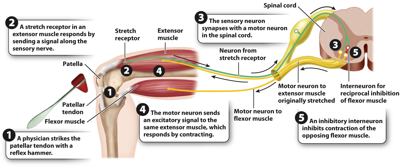
Let’s follow this reflex pathway. It starts with specialized “stretch” receptors located on the dendrites of a sensory neuron in the extensor muscles of the leg. These dendrites extend to cell bodies in ganglia alongside the spinal cord. These cell bodies have axons that extend into the spinal cord. The stretch receptors sense the stretch of the muscle that occurs during movement or in response to a physician’s strike of a reflex hammer. In response to a stretch, a signal is sent from the stretch receptor, through dendrite and cell body to the axon. In the spinal cord, the axon of the sensory neuron forms synapses with motor neurons that travel from the spinal cord back to the muscle where the stretch originated. The signal from the muscle stretch receptor stimulates the motor neurons to increase the activation of the muscle: the muscle contracts, and the leg extends at the knee.
This reflex arc does not include an interneuron: It is composed of just two neurons and one synapse. Transmission is delayed by communication at synapses, so the muscle contracts quickly because only one synapse is required to relay the sensory information back to the muscle.
As well as being a useful medical test, the knee-
Because muscles can only contract, opposing movements at a joint such as flexion and extension require flexor and extensor muscles on either side of the joint (Chapter 37). These are often activated out of phase (at different times) so that, when one set of muscles is activated (contracting), the other is inhibited (relaxed). The alternating extension and flexion of a leg during walking and running provide an example. This pattern of joint and limb movement is achieved by reciprocal inhibition: When stretch receptors of the knee extensor muscles are activated to stimulate these muscles to extend the knee, they also inhibit the activity of opposing muscles that flex the knee (Fig. 35.19).
Reciprocal inhibition of opposing sets of muscles occurs in the spinal cord. Axons of the stretch receptor neurons not only synapse with motor neurons of extensor muscles, but also synapse with inhibitory interneurons that inhibit motor neuron stimulation of the opposing flexor muscles. This inhibitory reflex pathway contains two synapses, one between the sensory neuron and interneuron and the second between the interneuron and the motor neuron to the flexor muscle.
Quick Check 5 Why is an interneuron needed to provide reciprocal inhibition of the flexor muscle when an extensor muscle is activated?
Quick Check 5 Answer
The sensory neuron releases only one type of neurotransmitter, which is excitatory for the extensor muscle from which the stretch receptor was stimulated. An interneuron provides an inhibitory synapse to the flexor muscle.
Reciprocal inhibition also operates between the right and left sides of the body. For example, reciprocal inhibition helps to control the movement of the right and left limbs. In this case, interneurons cross the spinal cord to control the timing of activity by flexor and extensor muscles of the opposite limb. Reciprocal inhibition is also involved in the Mauthner cell circuit of fish to ensure that only one side of the fish’s body bends away from the stimulus to escape. The local spinal circuits that provide reciprocal inhibition are therefore fundamental to the alternating motion of the body and limbs that characterizes the movements of most animals.
In other cases, more complex circuits in the brain may act to coordinate sensory input and motor output. These circuits integrate other sources of sensory information, such as vision and balance, with conscious commands from the brain to voluntarily control motor behavior. In the next chapter, we look more closely at these sensory circuits.