Hair cells detect the physical vibrations of sound.
What our ears interpret as sound are alternating waves of pressure traveling through the air. The frequency of the waves—
Insects hear by sensing airborne vibrations with small hairs on their antennae or other regions of their body. Hairs of different lengths detect different frequencies because the shorter hairs are stiffer than the longer hairs and vibrate at higher frequencies. Frequency is measured in hertz (Hz), or cycles per second. Male mosquitoes have small antennal hairs that vibrate in response to the 500-
Many insects also have specialized ears that sense sound by means of a tympanic membrane. The tympanic membrane is a thin sheet of tissue at the surface of the ear that vibrates in response to sound waves, amplifying airborne vibrations. The vibration excites hair cells attached to the inside of the tympanic membrane, and these hair cells in turn excite other neurons to fire action potentials. Crickets and other singing insects use their ears to sense the chirping or buzzing of potential mates.
Hearing is most widespread and developed in terrestrial vertebrates. The ears of amphibians, reptiles, and birds have an external tympanic membrane that vibrates when sound waves strike its surface. This membrane is similar to the tympanic membrane of insects but evolved independently and therefore is an example of convergent evolution. Terrestrial vertebrate ears can detect a broad range of sound frequencies. For humans, the range of audible frequencies is from 20 to 20,000 Hz. For dogs, the audible range is 70 to 44,000 Hz, and some rodents can hear up to 80,000 Hz. Birds generally hear in the same frequency range as mammals, which is one reason bird songs are pleasing to our ear. Frogs and reptiles generally hear at lower frequencies than mammals and birds. Vertebrate ears can also make fine distinctions between close frequencies and can detect softer sounds because external structures amplify the sounds before they reach the hair cells.
The ears of mammals have an external structure, the pinna, which enhances the reception of sound waves contacting the ear. Notice the heightened sensitivity that this structure provides for hearing when you cup your hands behind your ears. It is no coincidence that dogs and rabbits perk up their ears when alerted by a sound: This action orients them to the direction of the sound’s source.
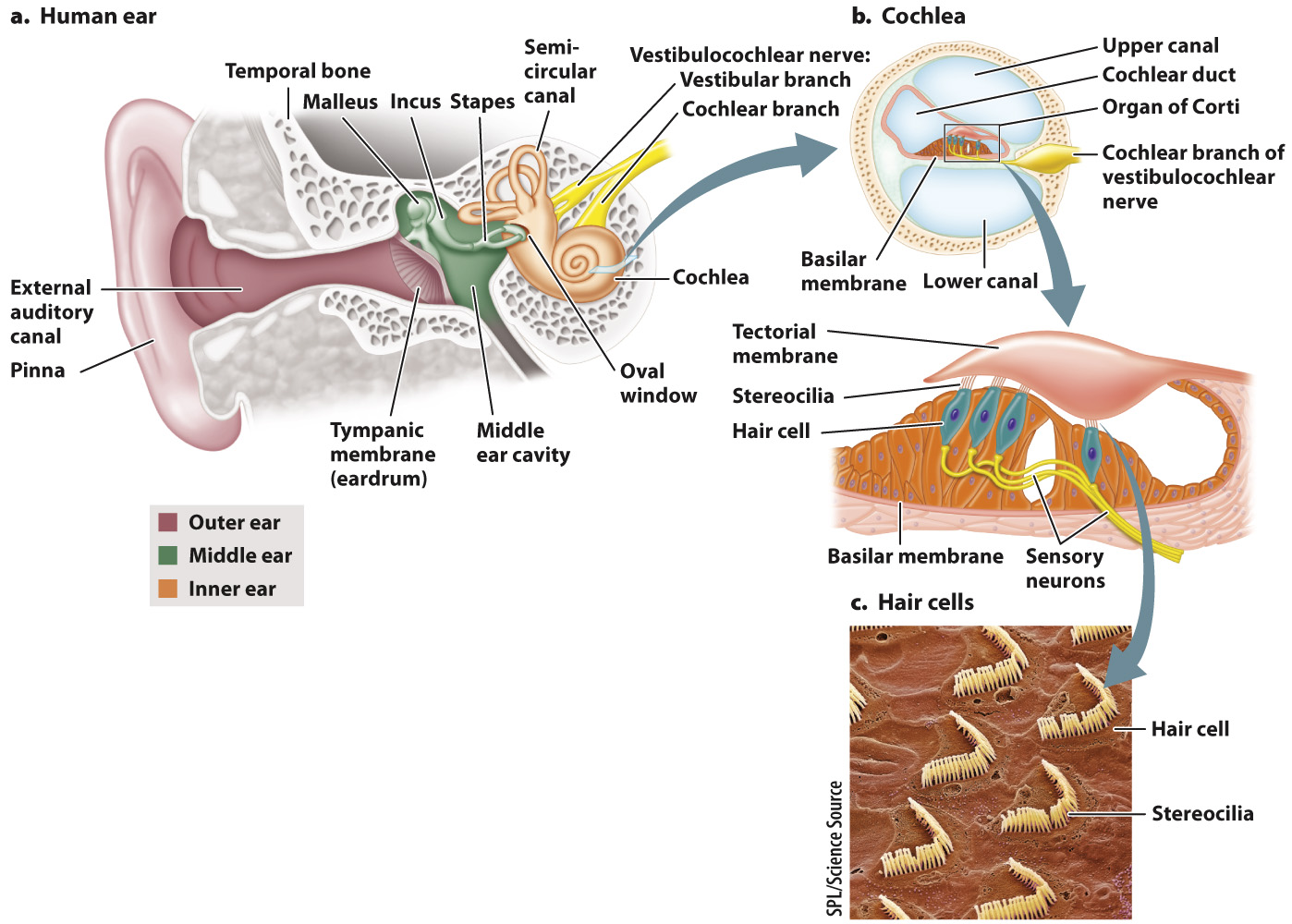
Fig. 36.8 shows the structure of the human ear. The pinna is part of the outer ear, which also includes the ear canal and tympanic membrane, or mammalian eardrum, which transmits airborne sounds into the ear. The middle ear contains three small bones, or ossicles, called the malleus, incus, and stapes, which amplify the waves that strike the tympanic membrane. The stapes connects to a thin membrane called the oval window of the cochlea in the inner ear. The cochlea (which means “snail” in Latin) is a coiled chamber within the skull that contains hair cells that convert pressure waves into an electrical impulse that is sent to the brain.
The mammalian eardrum evolved from the tympanic membrane of reptiles and earlier amphibians. The three middle ear bones are also evolutionarily related to three bones in reptiles, but only the stapes functions in reptiles to transmit vibrations of the tympanic membrane to the inner ear. In reptiles, the malleus and incus help support jaw movements during feeding. As mammals evolved from their reptilian ancestors, these two bones were incorporated with the stapes to form the middle ear bones that transmit sounds from the eardrum to the inner ear.
The process of hearing can be separated into three stages:
- Amplification. Sound vibrations received by the outer ear are transmitted by the eardrum and amplified by the three bones in the middle ear (Fig. 36.8a). Through piston-
like actions, the stapes transmits the energy of its movements to vibrations of the oval window of the cochlea in the inner ear. Vibrations transmitted from the eardrum through the middle ear to the oval window are amplified more than 30 times because of the larger size of the eardrum compared to the oval window and the lever- like action of the three bones. - Transfer of sound vibration to fluid pressure waves. The cochlea contains fluid and an upper and a lower canal separated by a basilar membrane and cochlear duct (Fig. 36.8b). Vibrations of the oval window cause fluid pressure waves in both canals at nearly the same time.
- Mechanoreception by hair cells within the cochlea. The cochlear duct houses the organ of Corti, which contains specialized hair cells with stereocilia. These hair cells are supported by the basilar membrane. The stereocilia of mammalian hair cells form a “V” and project into a rigid tectorial membrane that does not move (Fig. 36.8c).
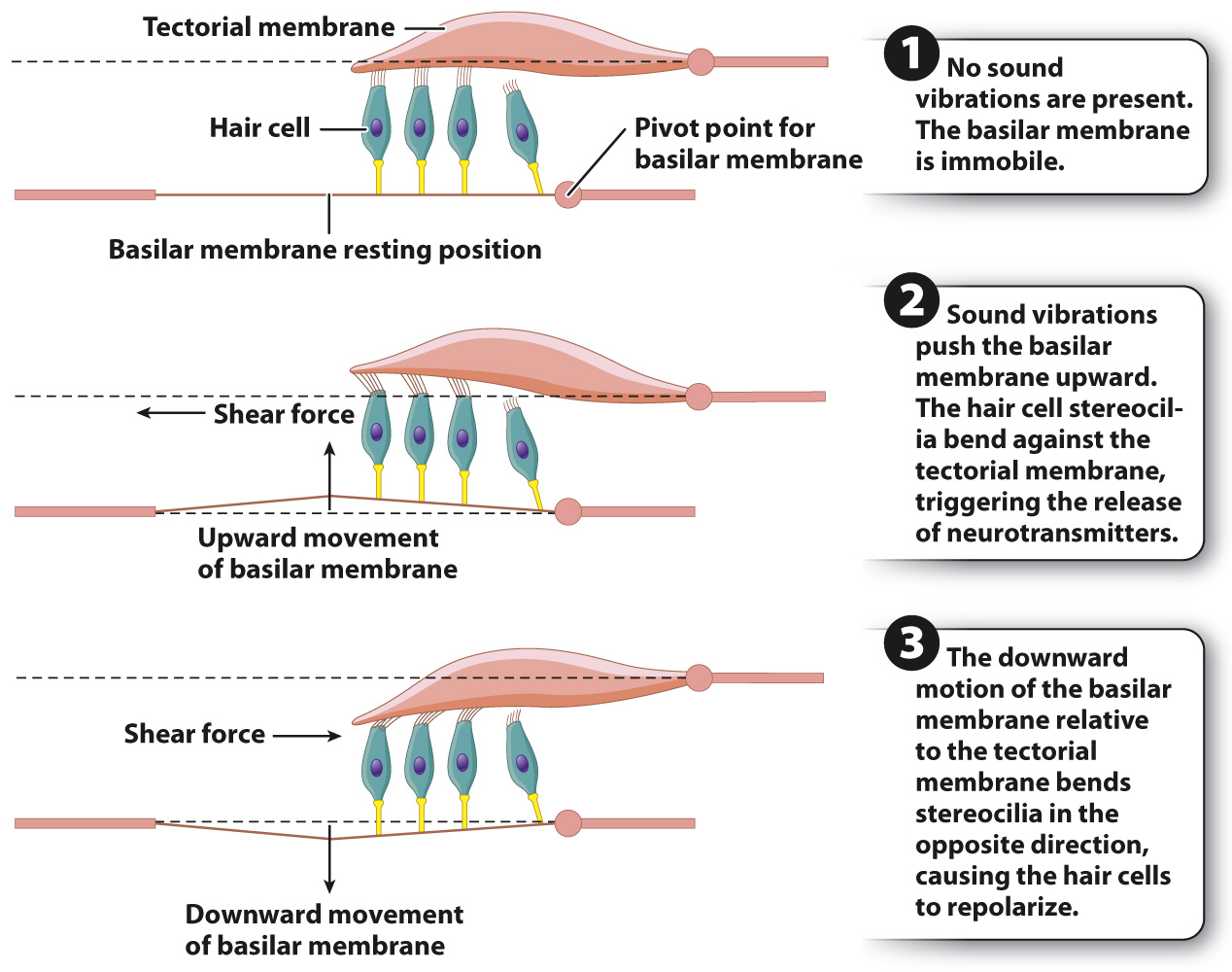
So far, we have seen pressure waves in the air transformed into vibrations of solid structures in the ear and then into waves of fluid. In the cochlea, motion is finally transduced into a nerve signal. The fluid vibrations within the cochlear canals induce motions of the basilar membrane relative to the tectorial membrane, bending the stereocilia of the hair cells back and forth in localized regions of the cochlea (Fig. 36.9). Bending of the stereocilia stimulates them to release excitatory neurotransmitters that cause postsynaptic neurons to fire action potentials. These are sensed as sound by neuronal networks in the auditory cortex, the area of the brain that processes sound.
Sound is characterized by amplitude (loudness) and frequency (pitch). Louder sounds produce larger fluid vibrations that cause the stereocilia to bend more, increasing their release of excitatory neurotransmitters. The release of more neurotransmitters increases the firing rate of the postsynaptic sensory neuron. The firing rate indicates intensity, or, in the case of hearing, loudness.
Many people can distinguish pure tones that differ by only a fraction of a percentage point in frequency. The ability to discriminate different sound frequencies is largely the result of differences in the mechanical properties of the basilar membrane along its length. At the apex, the basilar membrane is widest but thinnest and most flexible. At the base, the basilar membrane is narrow but thick and stiff. Thus, the basilar membrane is mechanically tuned to respond in different regions to different frequencies. Low-
Sound amplitude and frequency, together with vestibular sensing of gravity and head motion, are transmitted by the vestibulocochlear nerve (another cranial nerve) to the brain.