Color vision detects different wavelengths of light.
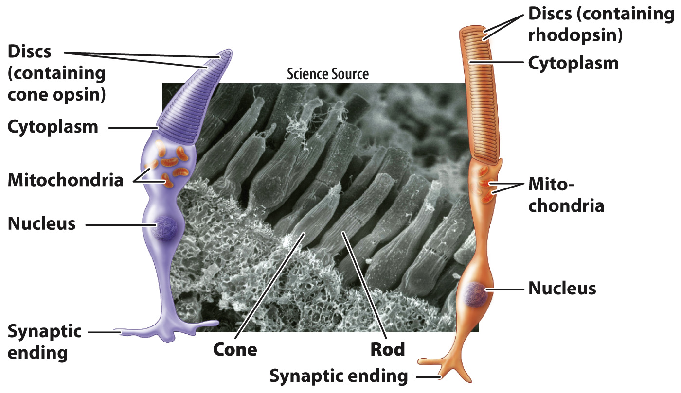
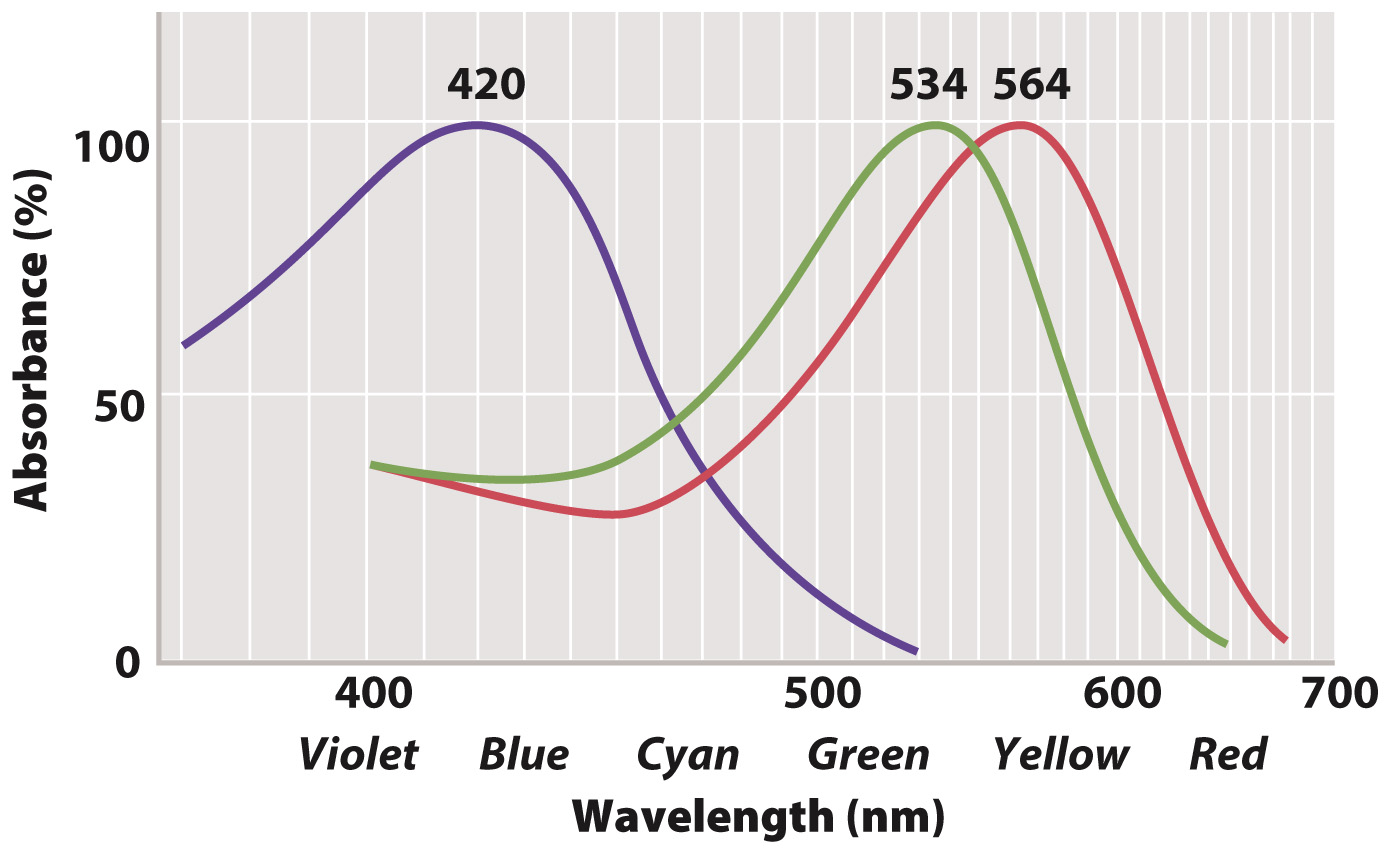
Color vision is crucial for many invertebrate and vertebrate animals. It is achieved by photoreceptor cells called cone cells that contain opsins sensitive to different wavelengths of light (Fig. 36.17). The more numerous rod cells are also sensitive to light, and most sensitive to blue-
Vertebrate cone cells likely evolved from a rod cell precursor. Most non-
Cone cells require higher levels of light than rod cells to become stimulated. The low sensitivity of cone cells to light makes it hard to detect color at night. Cone cells are most concentrated within the fovea of the retina, the center of the visual field of most vertebrates (see Fig. 36.15). Cone cells provide the sharpest vision. Animals with particularly sharp vision, such as hawks and other birds of prey, have an extremely concentrated number of cone cells in the fovea, approaching 1 million/mm2 (compared with about 150,000/mm2 in the human fovea). Birds of prey can see small prey on the ground from high in the air. Many birds also have eyes with two foveae, one projecting forward for binocular vision and one projecting to the side to enhance the sharpness of their side vision.
In contrast, rod cells are absent in the fovea but predominate in the periphery, making it easier to detect motion in the periphery, particularly at night. The eyes of many nocturnal predatory mammals, such as foxes and cats, contain mainly rod cells, enhancing nighttime vision. These animals also have a pigment located behind the retina that reflects light past the photoreceptors, enhancing the ability to sense light under low-
Quick Check 4 What is the primary role of rod cells, and what are the two roles of cone cells in the retina?
Quick Check 4 Answer
Rod cells are extremely sensitive to light, allowing an animal to see in dimly lit conditions. Cone cells provide color vision through opsins with absorption peaks at different light wavelengths, and they provide sharp vision.