The brain is divided into lobes with specialized functions.
The cerebral hemispheres are the largest structures of the mammalian brain (Fig. 36.23). They consist of a highly folded outer layer of gray matter about 4 mm thick that forms the cerebral cortex, which is made up of densely packed neuron cell bodies and their dendrites (Fig. 36.23a). The folds greatly increase the surface area of the cortex and are the result of selection for an increased number of cortical neurons within the limited volume of the skull. Deep inside the cerebral cortex is the white matter, which contains the axons of cortical neurons. The fatty myelin produced by glial cells surrounding the axon makes this region of the brain white. These axons are the means by which neurons in different regions of the gray matter communicate with neurons in other regions of the cortex and in deeper regions of the forebrain, midbrain, and hindbrain.
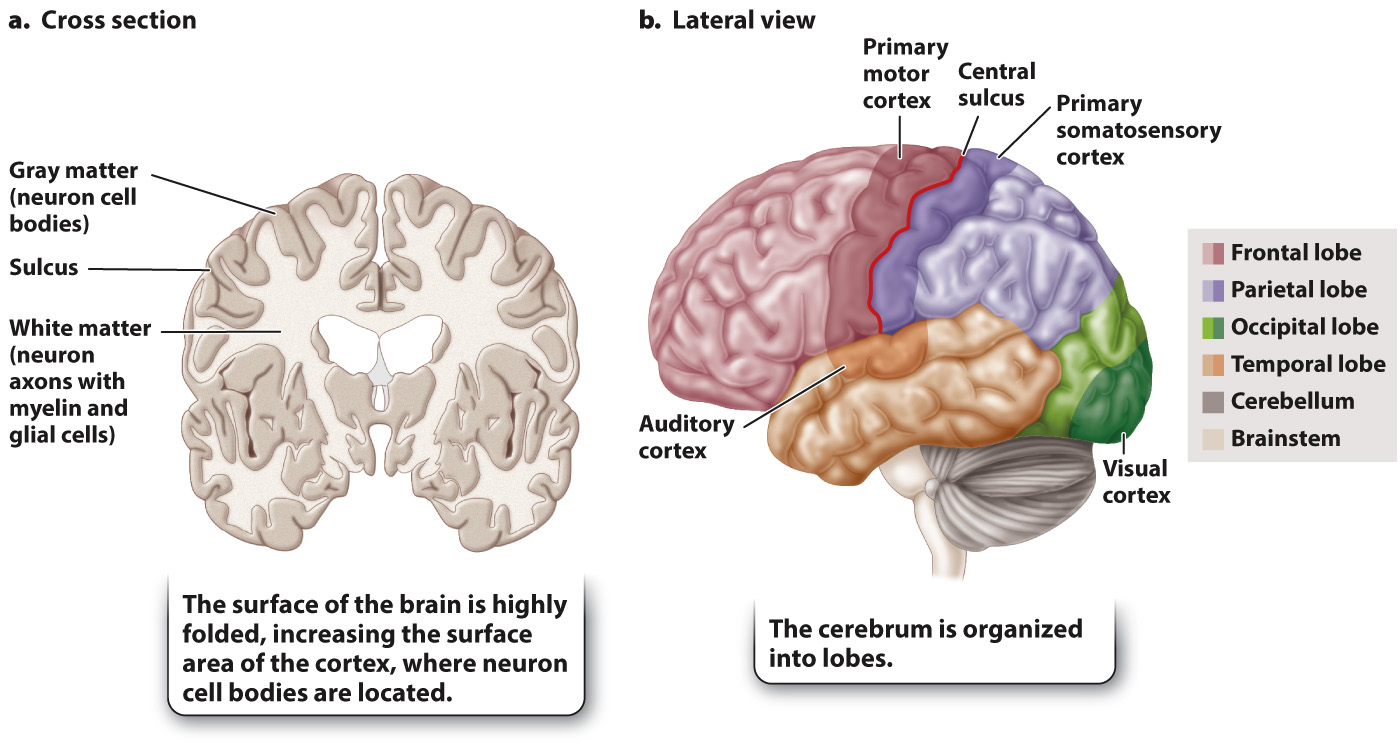
Major regions of the cerebral hemispheres are defined by clearly visible anatomical lobes separated in most cases by deep crevices called sulci (singular, sulcus) (Fig. 36.23b). The frontal lobe is located in the anterior region of the cerebral cortex (behind your forehead). It is important in decision making and planning. The parietal lobe is located posterior to the frontal lobe and is separated from the frontal lobe by the central sulcus. The parietal lobe controls body awareness and the ability to perform complex tasks, such as dressing. The temporal lobe lies below the parietal lobe. It is involved in processing sound, as well as performing other functions discussed below. Located behind the parietal lobe, at the back of the brain, is the occipital lobe, which processes visual information.
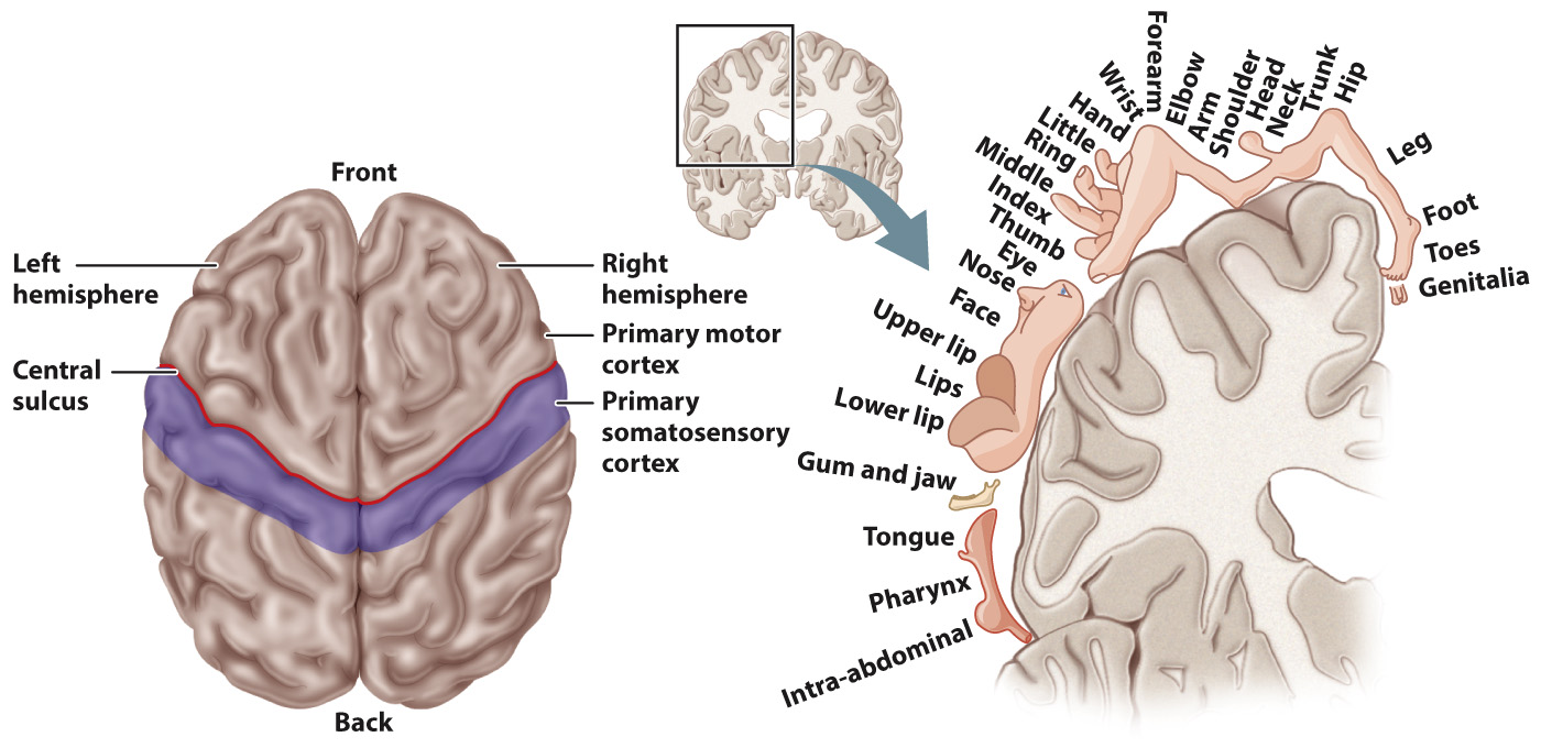
The central sulcus separates the primary motor cortex of the frontal lobe from the primary somatosensory cortex of the parietal lobe (Fig. 36.24). “Command” neurons in the primary motor cortex produce complex coordinated behaviors by controlling skeletal muscle movements. The primary somatosensory cortex integrates tactile information from specific body regions and relays this information to the motor cortex. Both cortices make connections to the opposite side of the body by sending axons that cross over in the brainstem and spinal cord. As a result, the right cortex controls the left side of the body and the left cortex controls the right side of the body.