Muscles contract by the sliding of myosin and actin filaments.

Using high-resolution light microscopy, two independent teams (physiologists Hugh Huxley and Jean Hanson in the United States and Andrew Huxley and Rolf Niedergerke in England) were able to quantify the changing banding pattern of sarcomeres when rabbit or frog myofibrils were stimulated to contract to different lengths. Their results led them to hypothesize that muscles produce force and change length by the sliding of actin filaments relative to myosin filaments. Their theory is known as the sliding filament model of muscle contraction.

The banding patterns of sarcomeres revealed the extent of overlap between actin and myosin filaments. When myofibrils contracted to short lengths, the sarcomeres were observed to have increased actin–myosin overlap. When myofibrils were stretched to longer lengths, actin–myosin overlap decreased (Fig. 37.6). However, the lengths of the myosin and actin filaments never varied. Consequently, nearly all of the length change during a muscle contraction results from the sliding of actin filaments with respect to myosin filaments within individual sarcomeres. The length change of the whole muscle fiber is therefore a sum of the fractions by which each sarcomere shortens along the fiber’s length.

image
FIG. 37.6 Sliding filament model. Muscle contraction, or shortening, results from the sliding of actin thin filaments relative to myosin thick filaments. Filament sliding changes the sarcomere banding pattern between Z discs.

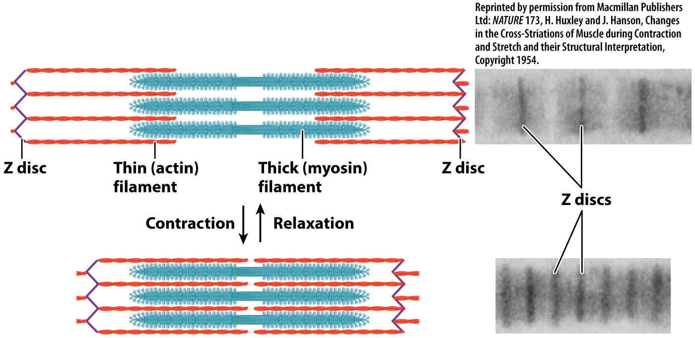
A muscle’s ability to generate force and change length is largely determined by the properties of its sarcomeres. Whereas sarcomere length is quite uniform in vertebrate animals (about 2.3 µm at rest), it is variable in invertebrate animals (ranging from as short as 1.3 µm to as long as 43 µm). Longer sarcomeres allow a greater degree of shortening. Thus, length changes are more limited in vertebrate muscles compared to those in some invertebrate muscles with long sarcomeres. For example, when the sarcomeres in an octopus tentacle contract, they can produce large motions of the tentacle. However, because vertebrates have shorter sarcomeres than an octopus, vertebrate muscles can shorten to produce movements more quickly.

Interactions between the myosin and actin filaments are what cause a muscle fiber to shorten and produce force. Along the myosin filament, one of the two heads of a myosin molecule at a given point in time binds to actin at a specific site to form cross-bridges between the myosin and actin filaments. The myosin filaments pull the actin filaments toward each other by means of these cross-bridges. The key to making the filaments slide relative to each other is the ability of the myosin head to undergo a conformational change and therefore pivot back and forth. The myosin head attaches to the actin filament and pivots forward, sliding the actin filament toward the middle of the sarcomere. The myosin head then detaches from the actin filament, pivots back, reattaches, and the cycle repeats. The movement of the myosin head is powered by ATP. Together, these events describe the cross-bridge cycle.

790

The cross-bridge cycle is just that—a cycle (Fig. 37.7). Let’s look at it in more detail:

image
FIG. 37.7 Cross-bridge cycle. The myosin head binds to actin and uses the energy of ATP to pull on the actin filament, causing muscle shortening.
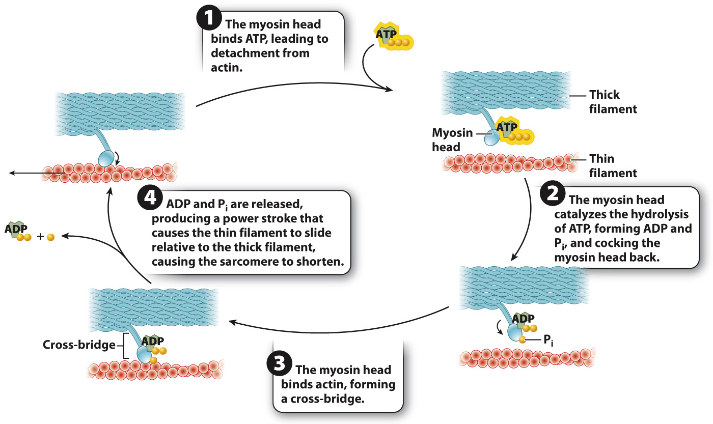
  1. The myosin head binds ATP. Binding of ATP allows the myosin head to detach from actin and readies it for attachment to actin.

  2. The myosin head hydrolyzes ATP to ADP and inorganic phosphate (Pi). Hydrolysis of ATP results in a conformational change in which the myosin head is cocked back. ADP and Pi remain bound to the myosin head. Because ADP and Pi are bound rather than released, the myosin head is in a high-energy state.

  3. The myosin head then binds actin, forming a cross-bridge.

  4. When the myosin head binds actin, the myosin head releases ADP and Pi. The result is another conformational change in the myosin head, called the power stroke. During the power stroke, the myosin head pivots forward and generates a force, causing the myosin and actin filaments to slide relative to each other over a distance of approximately 7 nm. The power stroke pulls the actin filaments toward the sarcomere midline.

The myosin head then binds a new ATP molecule (step 1 again), allowing it to detach from actin, and the cycle repeats. After detachment, the myosin head again hydrolyzes ATP, allowing it to return to its original conformation and bind to a new site farther along the actin filament. With the release of ADP and Pi, the myosin head undergoes another cycle of force generation and movement.

Individual muscle contractions are the result of many successive cycles of cross-bridge formation and detachment. During these cycles, muscle cells convert the chemical energy released by ATP into force and the kinetic energy of movement. Muscle fibers that contract especially quickly, such as those that power insect wing flapping or produce the sound of cicadas and rattlesnake tails, express myosin molecules that have high rates of ATP hydrolysis, allowing faster rates of cross-bridge cycling, force development, and shortening. Thus, myosin functions as both a structural protein and an enzyme.

Because each thick filament can interact with as many as six actin filaments, at any one time numerous cross-bridges anchor the lattice of myosin filaments, while other myosin heads are detached to find new binding sites. When summed over millions of cross-bridges, these molecular events generate the force that shortens the whole muscle.

791

Quick Check 1 When an animal dies, its limbs and body become stiff because its muscles go into rigor mortis (literally, rigor mortis means “stiffness of death”). Why would the loss of ATP following death cause this to happen?

Quick Check 1 Answer

Without newly synthesized ATP, myosin cross-bridges formed with actin cannot detach from their actin binding sites, so they remain in the bound state and make the muscle stiff.