The proximal convoluted tubule reabsorbs solutes by active transport.
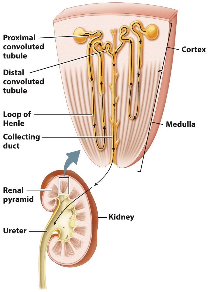
The renal tubule of the mammalian kidney is divided into three sections—
The filtrate initially moves into the first portion of the renal tubule, the proximal convoluted tubule (Fig. 41.17). Here, electrolytes and other nutrients that the animal requires are reabsorbed into the blood. To maximize absorption, the proximal convoluted tubule has thick walls composed of epithelial cells with fingerlike projections called microvilli on their surfaces, similar to those found in the small intestine (Chapter 40). The microvilli form a brush border along the inside (lumen) of the tubule, greatly increasing the cells’ surface area for solute and water reabsorption. The epithelial cells also possess numerous mitochondria that produce the ATP needed to actively reabsorb solutes from the filtrate into the bloodstream.
The proximal tubule reabsorbs all the glucose and amino acids filtered by the glomerulus, as well as most sodium and chloride ions. Because the proximal tubule is permeable to water, water diffuses by osmosis in the same direction as the electrolytes and solutes. By the time the filtrate leaves the proximal convoluted tubule, about 75% of the water, along with most of the electrolytes and solutes, has been reabsorbed into the bloodstream. What’s left is primarily water, urea, and electrolytes.