The female reproductive system produces eggs and supports the developing embryo.
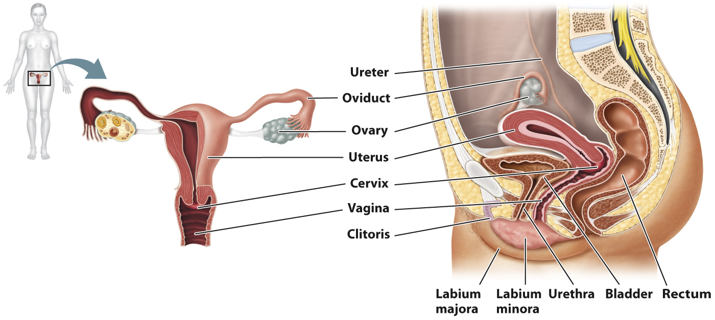
The female reproductive system, like the male reproductive system, produces gametes, but is also specialized to support the developing embryo. Developing female gametes, called oocytes, which mature into ova, are produced in the two female gonads, called ovaries (Fig. 42.14), and are released monthly in response to hormones in the process of menstruation, discussed in the next section.
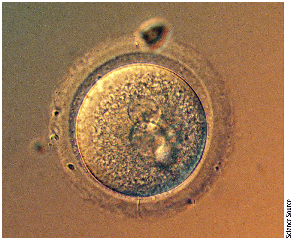
The oocyte is a remarkable and unique cell (Fig. 42.15). In contrast to sperm, which are small and motile, the oocyte is the largest cell by volume in the human body and is visible to the naked eye; it is about the size of the period at the end of this sentence. Its large size reflects the presence of large amounts of cytoplasm containing molecules that direct the early cell divisions following fertilization and food supply in the form of yolk. Whereas a male produces millions of sperm, a female usually produces just one oocyte each month, as she will nourish and support the developing embryo if the oocyte is fertilized.
Following release from the ovary, the oocyte travels through the fallopian tube, or oviduct (see Fig. 42.14). There are two fallopian tubes, one for each ovary. The fallopian tubes have featherlike projections on their ends that help channel the egg into the tube and not into the abdominal cavity.
The oocyte travels from the fallopian tube to the uterus, or womb (see Fig. 42.14). The uterus is a hollow organ with thick, muscular walls that is adapted to support the developing embryo if fertilization occurs and to deliver the baby during birth. In humans, the uterus is usually a single pear-
The end of the uterus is called the cervix, or neck, of the uterus (see Fig. 42.14). Under the influence of hormones, the cervix produces different kinds of mucus capable of either blocking or guiding sperm through the cervix.
The uterus is continuous with the vagina, or birth canal (see Fig. 42.14). The vagina is a tubular channel connecting the uterus to the exterior of the body. It is highly elastic, allowing it to stretch during sexual intercourse and birth.
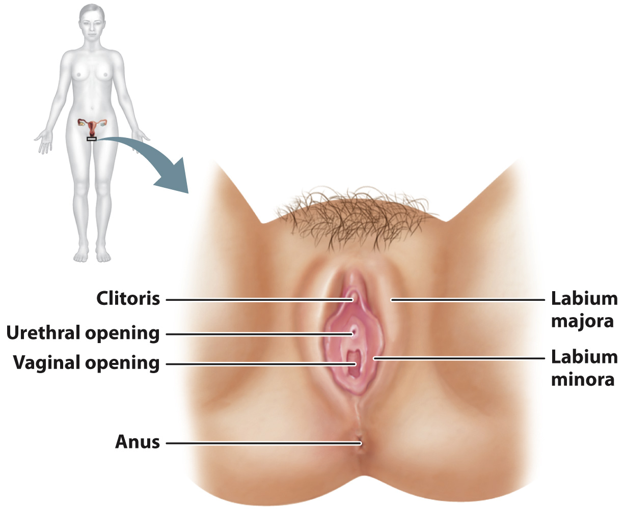
The external genitalia of the female are collectively called the vulva (Fig. 42.16). The vulva includes two folds of skin, the labia majora and labia minora. The labia minora meet at the clitoris, the female homolog of the glans penis.