T cells have T cell receptors on their surface that recognize an antigen in association with MHC proteins.
All T cells, like B cells, originate in the mammalian bone marrow. However, unlike B cells, T cells mature in the thymus. A mature T cell is characterized by the presence on the plasma membrane of a T cell receptor (TCR), a protein receptor that recognizes and binds to the antigen.
In many ways, TCRs are similar to antibodies on the surface of B cells. For example, TCRs recognize antigens with a specific structure. In addition, there is a great diversity of TCRs that differ from one another, but each T cell has just one type of TCR on its surface. Binding of TCR to an antigen triggers the T cell to divide into clones, resulting in a pool of T cells that are each specific for a given antigen. Finally, like antibodies, the diversity of TCRs among different T cells results from genomic rearrangement of V, D, J, and C gene segments.
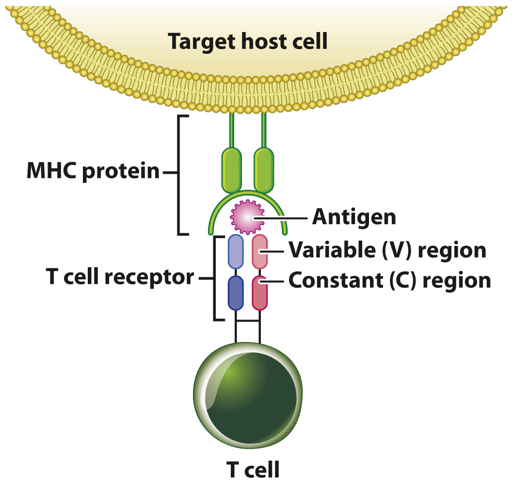
However, TCRs are different from antibodies in important ways. First, they are composed of two, rather than four, polypeptide chains (Fig. 43.15). Second, they are not secreted like antibodies, but are always membrane-
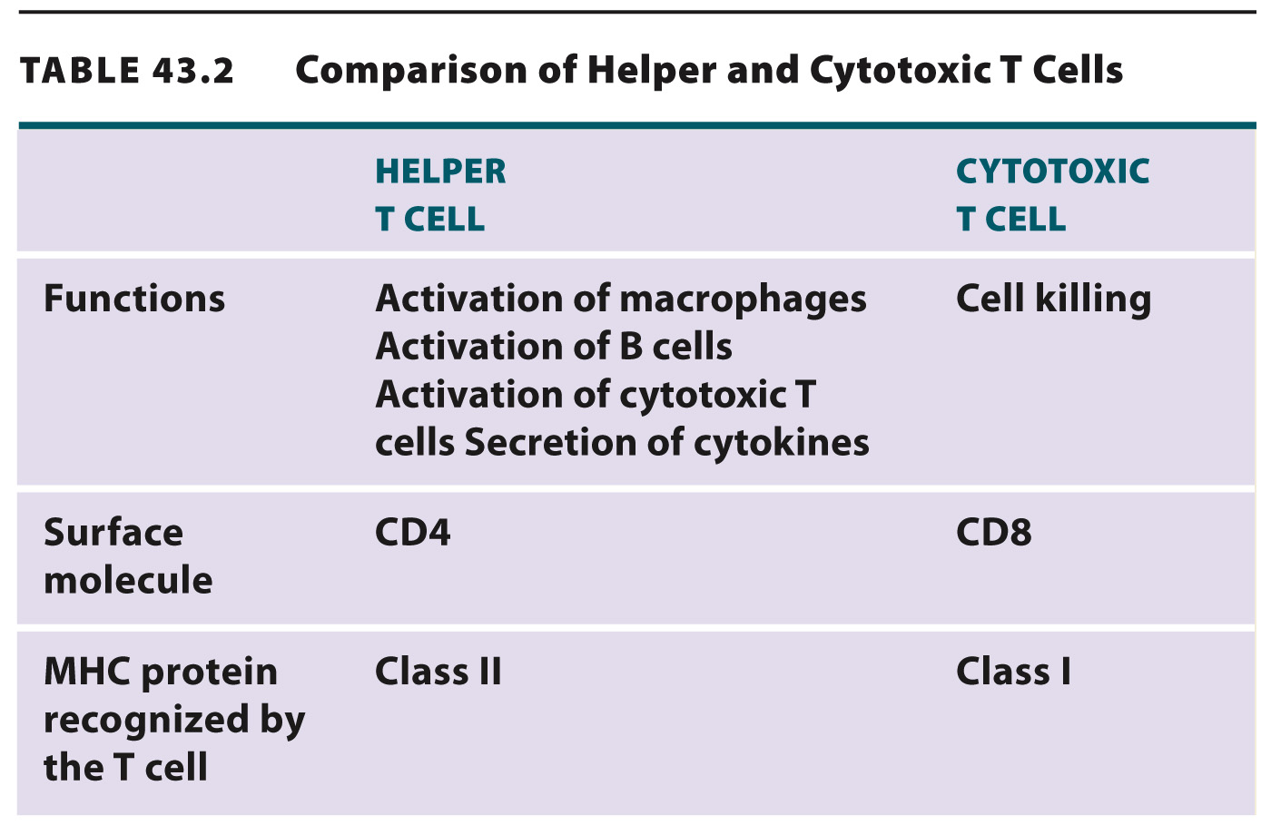
The MHC is a cluster of genes present in all mammals that encode proteins on the surface of cells. The MHC is composed of many genes with a high rate of polymorphism, meaning that there is a lot of variation in the gene sequence (and consequently in the protein sequence) among different individuals. In humans and mice, the genes are divided into three classes: Class I genes are expressed on the surface of all nucleated cells; class II genes are expressed on the surface of macrophages, dendritic cells, and B cells; and class III genes encode several proteins of the complement system and proteins involved in inflammation.
How do helper and cytotoxic T cells recognize and respond to antigens? Let’s consider the activation of helper T cells first (Fig. 43.16). When an antigen enters the body, it may be recognized by an antibody directly or be taken up by antigen-
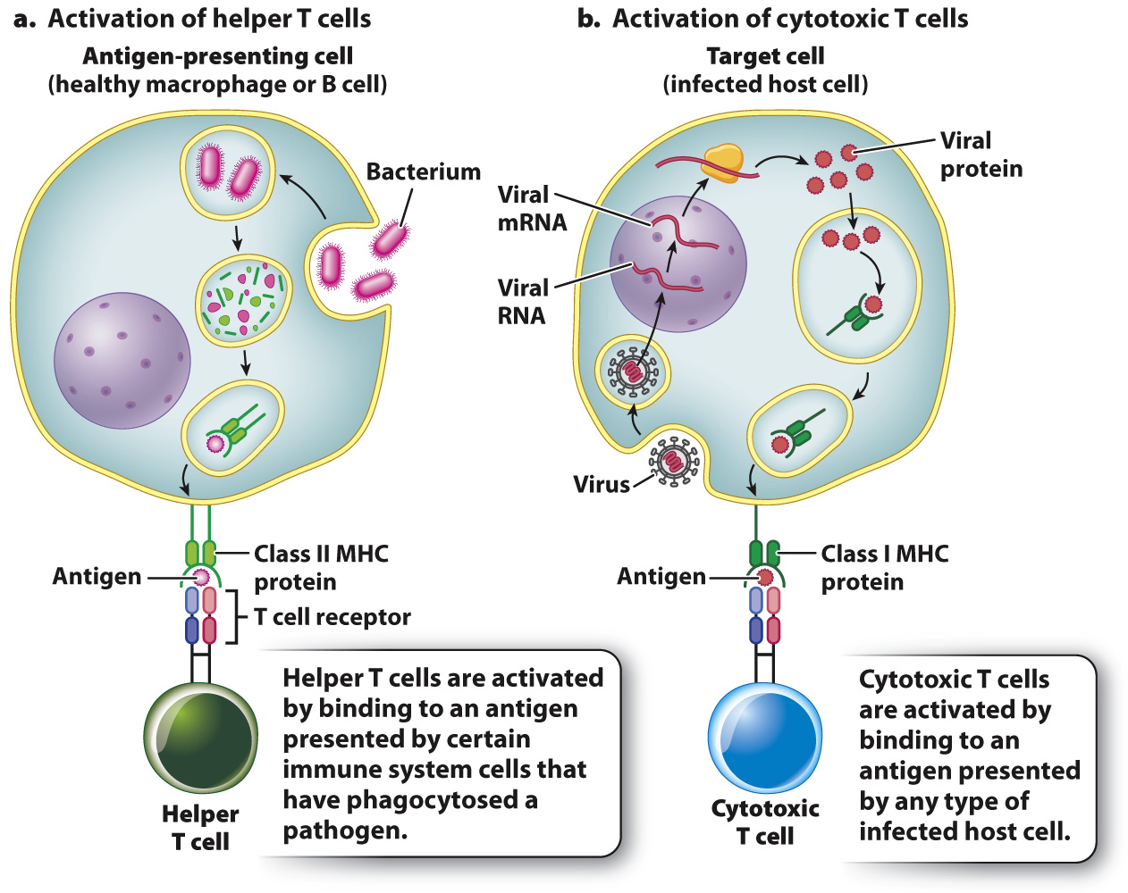
Cytotoxic T cells also recognize antigen displayed by host cells, but only antigens that are associated with MHC class I proteins (Fig. 43.16). Because class I proteins are present on virtually all cells, cytotoxic T cells recognize and kill any host cell that becomes abnormal in some way. For example, a virus-
Once activated, T cells divide and form clones of helper or cytotoxic T cells. Some cells of each type are memory cells that provide long-
Quick Check 4 How does T cell activation differ from B cell activation?
Quick Check 4 Answer
T cells are activated when their surface T cell receptors bind to an antigen in association with an MHC molecule. By contrast, B cells are activated when their surface antibodies bind to a free antigen.