Signaling can occur over short distances.
Signaling can also occur between two cells that are close to each other (Fig. 9.4b). When cells are close to each other, they do not require a circulatory system to deliver the signaling molecule. Instead, the signaling molecule can simply move by diffusion between the two cells. This form of signaling is called paracrine signaling. In this case, signaling molecules travel distances of about 20 cell diameters, or a few hundred micrometers.
In paracrine signaling, the signal is usually a small, water-
HOW DO WE KNOW?
FIG. 9.5
Where do growth factors come from?
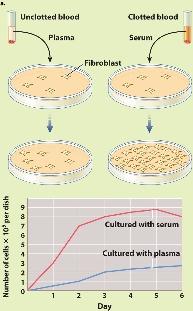
BACKGROUND Cells can be grown outside the body in culture. However, they survive and grow well only under certain conditions. Researchers hypothesized that there are substances that are required for cells to grow in culture, but the identity and source of these substances were for a long time unknown. A key insight came from the observation that chicken cells grow much better if they are cultured in the presence of blood serum rather than blood plasma. Blood serum is the liquid component of blood that is collected after blood has been allowed to clot. Blood plasma is also the liquid component of blood, but it is collected from blood that has not clotted. American biologists Nancy Kohler and Allan Lipton were interested in identifying the source of the factor in blood serum that allows cells to survive in culture.
HYPOTHESIS Since Kohler and Lipton knew that clotting depends on the release of substances from platelets, they hypothesized that a growth-
EXPERIMENT 1 The investigators first confirmed earlier observations using cells that are easily grown in culture called fibroblasts, which they obtained from mice. They cultured two sets of fibroblasts in small plastic dishes. To one of the cultures they added serum; to the other culture they added plasma. Then they monitored the rate of cell division in both culture dishes over time.
RESULTS 1 They observed that the rate of cell division in the fibroblasts cultured in serum was far greater than that of the cells cultured in plasma, as expected based on earlier experiments (Fig. 9.5a).
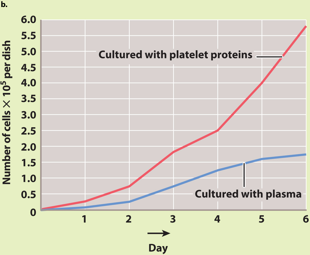
EXPERIMENT 2 To see if the factor is released directly from platelets, they prepared a solution of proteins made from purified platelets, added these proteins to cultured fibroblasts, and measured cell growth.
RESULTS 2 They found that the solution of platelet proteins also caused the growth of fibroblasts to increase compared to the growth of fibroblasts in plasma (Fig. 9.5b).
CONCLUSION Kohler and Lipton concluded that the growth-
FOLLOW-
SOURCE Kohler, N., and A. Lipton. 1974. “Platelets as a Source of Fibroblast Growth-
Growth factors secreted by cells in an embryo work over short distances to influence the kind of cells their neighbors will become. In this way, they help shape the structure of the adult’s tissues, organs, and limbs. For example, in developing vertebrates, paracrine signaling by the growth factor Sonic Hedgehog (yes, it’s named after a video game character) ensures that the motor neurons in your spinal cord are located properly, that the bones of your vertebral column form correctly, and that your thumb and pinky fingers are on the correct sides of your hands.
A specialized form of short-
In some cases, signaling molecules may be released by a cell and then bind to receptors on the very same individual cell. Such cases, where signaling cell and responding cell are one and the same, are examples of autocrine signaling (Fig. 9.4c). Autocrine signaling is especially important to multicellular organisms during the development of the embryo (Chapters 20 and 42). For example, once a cell differentiates into a specialized cell type, autocrine signaling is sometimes used to maintain this developmental decision. In addition, autocrine signaling can be used by cancer cells to promote cell division.