2.3 The Cerebral Cortex and Our Divided Brain
Please proceed to the next section.
The Cerebral Cortex
2-
The people who first dissected and labeled the brain used the language of scholars—Latin and Greek. Their words are actually attempts at graphic description: For example, cortex means “bark,” cerebellum is “little brain,” and thalamus is “inner chamber.”
cerebral [seh-REE-bruhl] cortex the intricate fabric of interconnected neural cells covering the cerebral hemispheres; the body’s ultimate control and information-processing center.
Older brain networks sustain basic life functions and enable memory, emotions, and basic drives. Newer neural networks within the cerebrum—the two cerebral hemispheres contributing 85 percent of the brain’s weight—
As we move up the ladder of animal life, the cerebral cortex expands, tight genetic controls relax, and the organism’s adaptability increases. Frogs and other small-
RETRIEVAL PRACTICE
- Which area of the human brain is most similar to that of less complex animals? Which part of the human brain distinguishes us most from less complex animals?
The brainstem; the cerebral cortex
75
Structure of the Cortex
If you opened a human skull, exposing the brain, you would see a wrinkled organ, shaped somewhat like the meat of an oversized walnut. Without these wrinkles, a flattened cerebral cortex would require triple the area—
Each hemisphere’s cortex is subdivided into four lobes, separated by prominent fissures, or folds (FIGURE 2.23). Starting at the front of your brain and moving over the top, there are the frontal lobes (behind your forehead), the parietal lobes (at the top and to the rear), and the occipital lobes (at the back of your head). Reversing direction and moving forward, just above your ears, you find the temporal lobes. Each of the four lobes carries out many functions, and many functions require the interplay of several lobes.
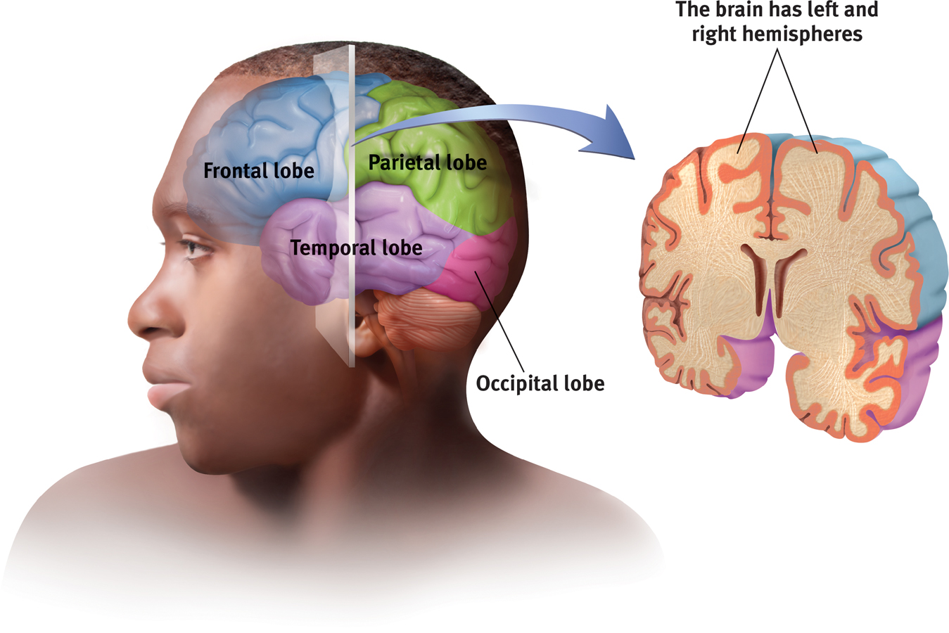
The cortex and its basic subdivisions
frontal lobes portion of the cerebral cortex lying just behind the forehead; involved in speaking and muscle movements and in making plans and judgments.
parietal [puh-RYE-uh-tuhl] lobes portion of the cerebral cortex lying at the top of the head and toward the rear; receives sensory input for touch and body position.
occipital [ahk-SIP-uh-tuhl] lobes portion of the cerebral cortex lying at the back of the head; includes areas that receive information from the visual fields.
temporal lobes portion of the cerebral cortex lying roughly above the ears; includes the auditory areas, each receiving information primarily from the opposite ear.
Functions of the Cortex
More than a century ago, surgeons found damaged cortical areas during autopsies of people who had been partially paralyzed or speechless. This rather crude evidence did not prove that specific parts of the cortex control complex functions like movement or speech. After all, if the entire cortex controlled speech and movement, damage to almost any area might produce the same effect. A TV with its power cord cut would go dead, but we would be fooling ourselves if we thought we had “localized” the picture in the cord.
motor cortex an area at the rear of the frontal lobes that controls voluntary movements.
Motor Functions Scientists had better luck in localizing simpler brain functions. For example, in 1870, German physicians Gustav Fritsch and Eduard Hitzig made an important discovery: Mild electrical stimulation to parts of an animal’s cortex made parts of its body move. The effects were selective: Stimulation caused movement only when applied to an arch-
MAPPING THE MOTOR CORTEX Lucky for brain surgeons and their patients, the brain has no sensory receptors. Knowing this, Otfrid Foerster and Wilder Penfield were able to map the motor cortex in hundreds of wide-
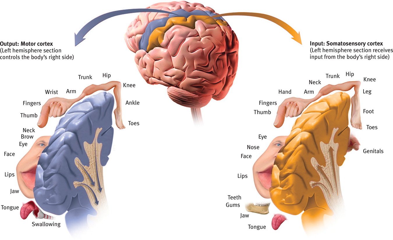
Left hemisphere tissue devoted to each body part in the motor cortex and the somatosensory cortex As you can see from this classic though inexact representation, the amount of cortex devoted to a body part in the motor cortex (in the frontal lobes) or in the somatosensory cortex (in the parietal lobes) is not proportional to that body part’s size. Rather, the brain devotes more tissue to sensitive areas and to areas requiring precise control. Thus, the fingers have a greater representation in the cortex than does the upper arm.
76
RETRIEVAL PRACTICE
- Try moving your right hand in a circular motion, as if cleaning a table. Then start your right foot doing the same motion, synchronized with your hand. Now reverse the right foot’s motion, but not the hand’s. Finally, try moving the left foot opposite to the right hand.
- Why is reversing the right foot’s motion so hard?
- Why is it easier to move the left foot opposite to the right hand?
1. The right limbs’ opposed activities interfere with each other because both are controlled by the same (left) side of your brain.
2. Opposite sides of your brain control your left and right limbs, so the reversed motion causes less interference.
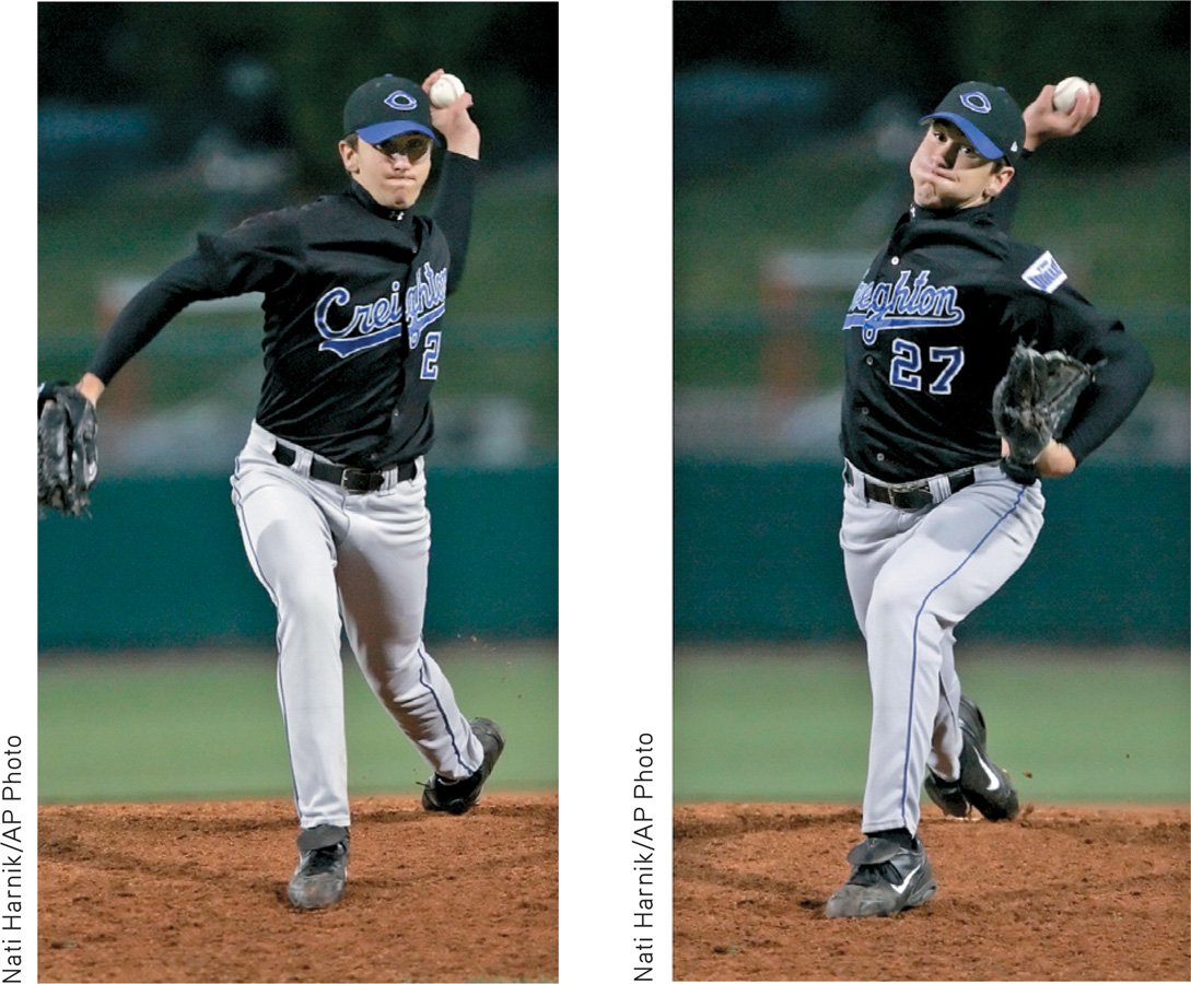
More recently, scientists were able to predict a monkey’s arm motion a tenth of a second before it moved—
77
BRAIN–
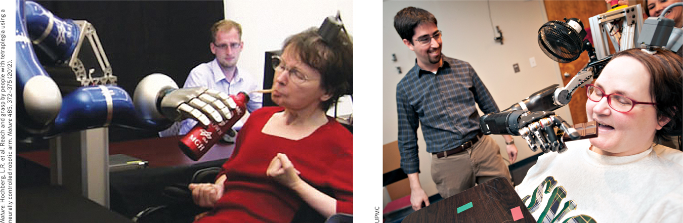
Mind over matter Strokes caused Cathy’s (left) complete paralysis, as did a neurodegenerative disease for Jan (right). Yet, thanks to a tiny, 96-
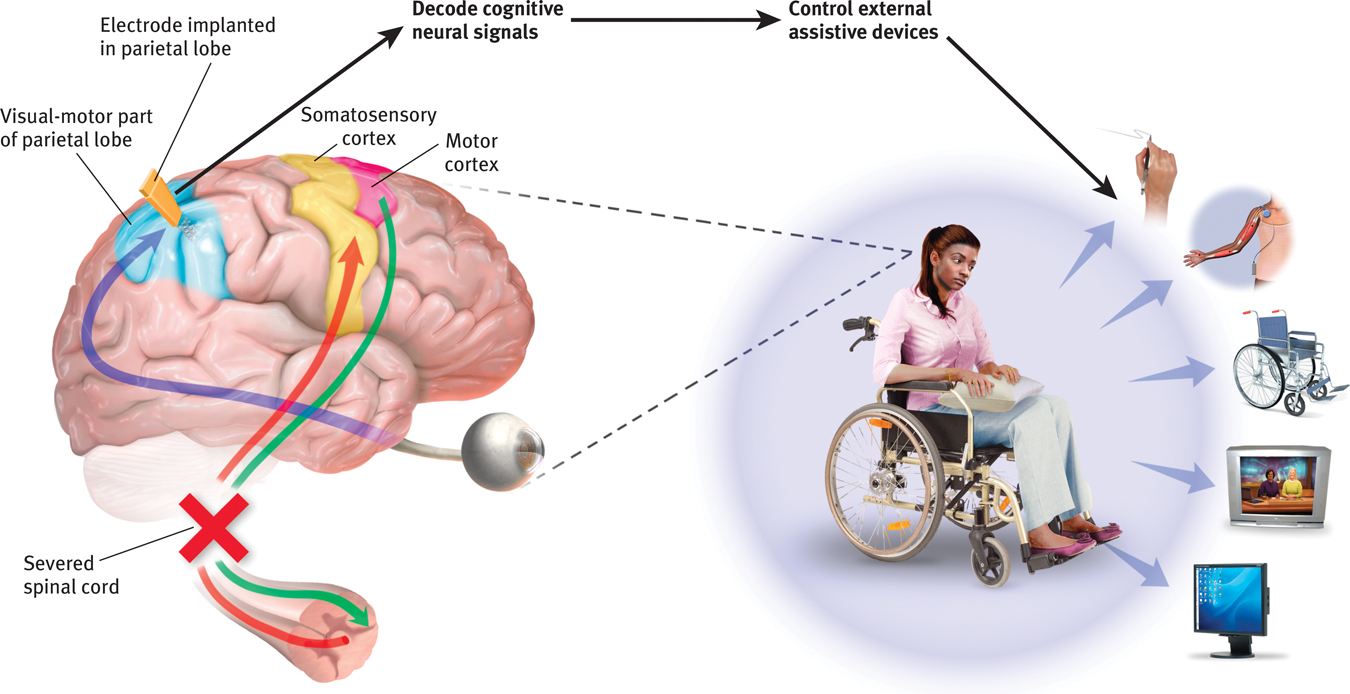
Research has also recorded messages not from the arm-
If this technique works, why not use it to capture the words a person can think but cannot say (for example, after a stroke)? Cal Tech neuroscientist Richard Andersen (2004, 2005) has speculated that researchers could implant electrodes in speech areas, then “ask a patient to think of different words and observe how the cells fire in different ways. So you build up your database, and then when the patient thinks of the word, you compare the signals with your database, and you can predict the words they’re thinking. Then you take this output and connect it to a speech synthesizer. This would be identical to what we’re doing for motor control.” With this goal in mind, the U.S. Army is investing $6.3 million in neuroscientists’ efforts to build a helmet that might read and transmit soldiers’ thoughts (Piore, 2011).
Clinical trials of such cognitive neural prosthetics are now under way with people who have suffered paralysis or amputation (Andersen et al., 2010; Nurmikko et al., 2010). The first patient, a paralyzed 25-
Brain–
78
somatosensory cortex area at the front of the parietal lobes that registers and processes body touch and movement sensations.
Sensory Functions If the motor cortex sends messages out to the body, where does the cortex receive incoming messages? Penfield identified a cortical area—
The more sensitive the body region, the larger the somatosensory cortex area devoted to it (Figure 2.24). Your supersensitive lips project to a larger brain area than do your toes, which is one reason we kiss with our lips rather than touch toes. Rats have a large area of the brain devoted to their whisker sensations, and owls to their hearing sensations.
Scientists have identified additional areas where the cortex receives input from senses other than touch. Any visual information you are receiving now is going to the visual cortex in your occipital lobes, at the back of your brain (FIGURE 2.27 and FIGURE 2.28). Stimulated in the occipital lobes, you might see flashes of light or dashes of color. (In a sense, we do have eyes in the back of our head!) Having lost much of his right occipital lobe to a tumor removal, a friend was blind to the left half of his field of vision. Visual information travels from the occipital lobes to other areas that specialize in tasks such as identifying words, detecting emotions, and recognizing faces.
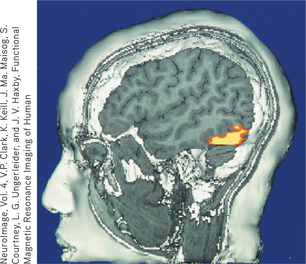
The brain in action This fMRI (functional MRI) scan shows the visual cortex in the occipital lobes activated (color represents increased bloodflow) as a research participant looks at a photo. When the person stops looking, the region instantly calms down.
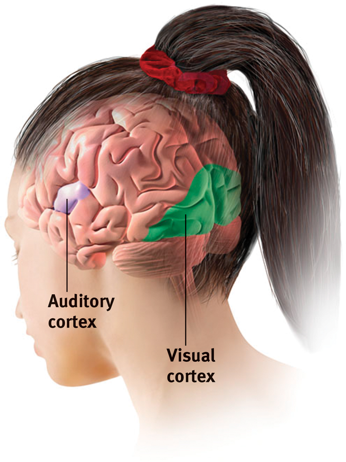
The visual cortex and auditory cortex The visual cortex in the occipital lobes at the rear of your brain receives input from your eyes. The auditory cortex, in your temporal lobes—
Any sound you now hear is processed by your auditory cortex in your temporal lobes (just above your ears; see Figure 2.28). Most of this auditory information travels a circuitous route from one ear to the auditory receiving area above your opposite ear. If stimulated in your auditory cortex, you might hear a sound. MRI scans of people with schizophrenia have revealed active auditory areas in the temporal lobes during the false sensory experience of auditory hallucinations (Lennox et al., 1999). Even the phantom ringing sound experienced by people with hearing loss is—
79
RETRIEVAL PRACTICE
- Our brain’s ______________ cortex registers and processes body touch and movement sensations. The ______________ cortex controls our voluntary movements.
somatosensory; motor
Association Areas So far, we have pointed out small cortical areas that either receive sensory input or direct muscular output. Together, these occupy about one-
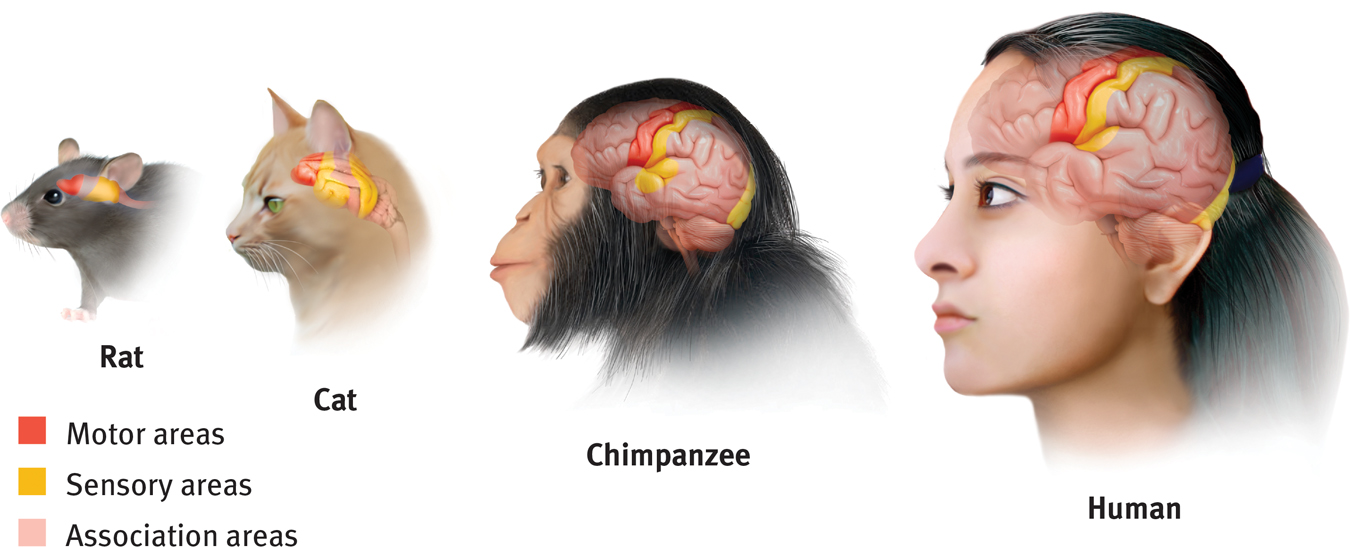
Areas of the cortex in four mammals More intelligent animals have increased “uncommitted” or association areas of the cortex. These vast areas of the brain are responsible for interpreting, integrating, and acting on sensory information and linking it with stored memories.
association areas areas of the cerebral cortex that are not involved in primary motor or sensory functions; rather, they are involved in higher mental functions such as learning, remembering, thinking, and speaking.
Electrically probing an association area won’t trigger any observable response. So, unlike the somatosensory and motor areas, association area functions cannot be neatly mapped. Their silence has led to what Donald McBurney (1996, p. 44) called “one of the hardiest weeds in the garden of psychology”: the claim that we ordinarily use only 10 percent of our brain. (If true, wouldn’t this imply a 90 percent chance that a bullet to your brain would strike an unused area?) Surgically lesioned animals and brain-
80
Association areas are found in all four lobes. The prefrontal cortex in the forward part of the frontal lobes enables judgment, planning, and processing of new memories. People with damaged frontal lobes may have intact memories, high scores on intelligence tests, and great cake-
Frontal lobe damage also can alter personality and remove a person’s inhibitions. Consider the classic case of railroad worker Phineas Gage. One afternoon in 1848, Gage, then 25 years old, was using a tamping iron to pack gunpowder into a rock. A spark ignited the gunpowder, shooting the rod up through his left cheek and out the top of his skull, leaving his frontal lobes damaged (FIGURE 2.30). The rod not only damaged some of Gage’s left frontal lobe’s neurons, but also about 11 percent of its axons that connect the frontal lobes with the rest of the brain (Van Horn et al., 2012). To everyone’s amazement, he was immediately able to sit up and speak, and after the wound healed he returned to work. But having lost some of the neural tracts that enabled his frontal lobes to control his emotions, the affable, soft-
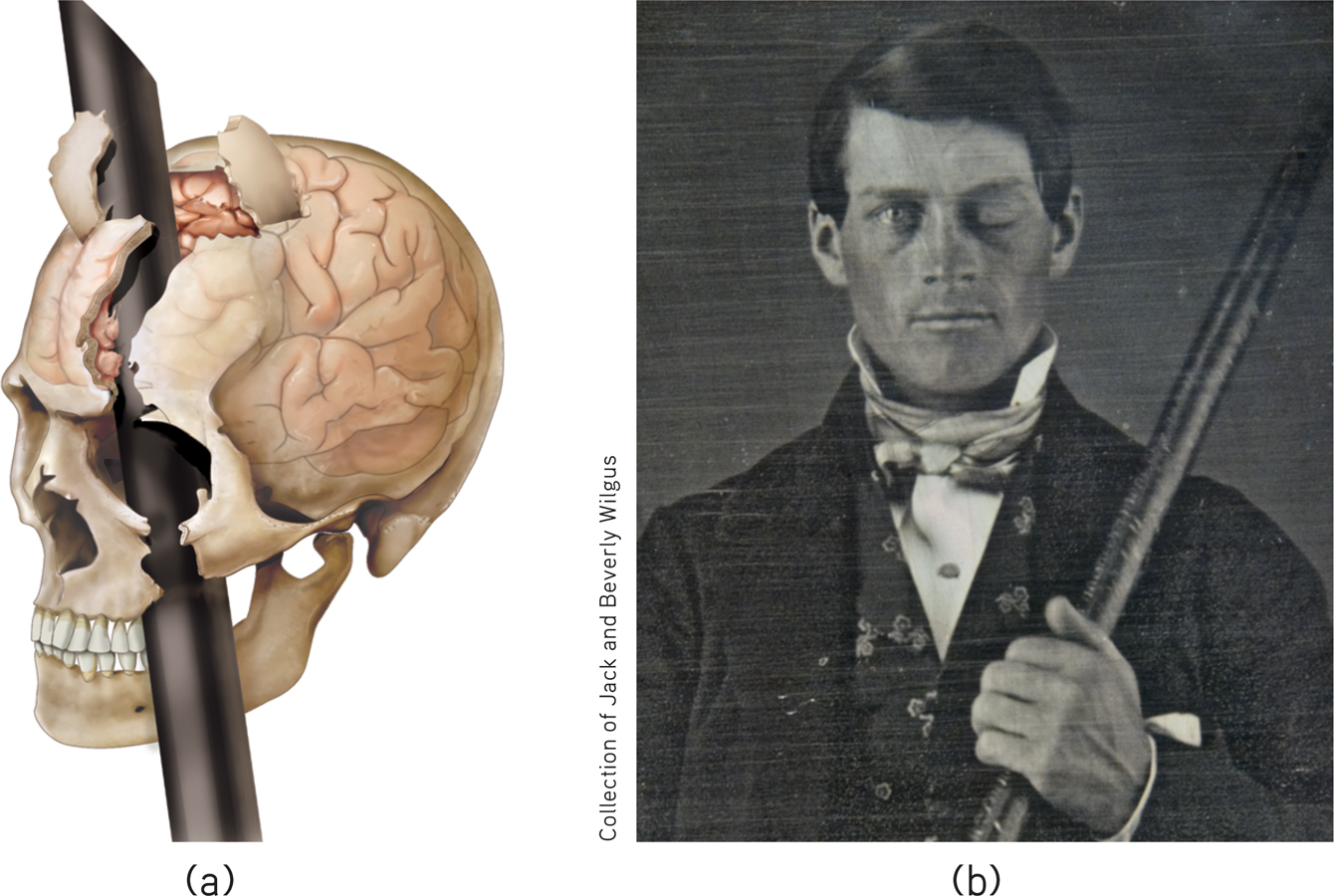
A blast from the past (a) Phineas Gage’s skull was kept as a medical record. Using measurements and modern neuroimaging techniques, researchers have reconstructed the probable path of the rod through Gage’s brain (Van Horn et al., 2012). (b) This photo shows Gage after his accident. (The image has been reversed to show the features correctly. Early photos, including this one, were actually mirror images.)
Studies of others with damaged frontal lobes have revealed similar impairments. Not only may they become less inhibited (without the frontal lobe brakes on their impulses), but their moral judgments may seem unrestrained by normal emotions. Would you advocate pushing one person in front of a runaway trolley to save five others? Most people do not, but those with damage to a brain area behind the eyes often do (Koenigs et al., 2007). With their frontal lobes ruptured, people’s moral compass seems to disconnect from their behavior.
Association areas also perform other mental functions. The parietal lobes, parts of which were large and unusually shaped in Einstein’s normal-
81
On the underside of the right temporal lobe, another association area enables us to recognize faces. If a stroke or head injury destroyed this area of your brain, you would still be able to describe facial features and to recognize someone’s gender and approximate age, yet be strangely unable to identify the person as, say, your grandmother.
Nevertheless, to reemphasize, we should be wary of using pictures of brain “hot spots” to create a new phrenology that locates complex functions in precise brain areas (Beck, 2010; Shimamura, 2010; Uttal, 2001). Complex mental functions don’t reside in any one place. There is no one spot in a rat’s small association cortex that, when damaged, will obliterate its ability to learn or remember a maze. Your memory, language, and attention result from the synchronized activity among distinct brain areas and neural networks (Knight, 2007). Ditto for religious experience. More than 40 distinct brain regions become active in different religious states, such as prayer and meditation, indicating that there is no simple “God spot” (Fingelkurts & Fingelkurts, 2009). The point to remember: Our mental experiences arise from coordinated brain activity.
RETRIEVAL PRACTICE
- Why are association areas important?
Association areas are involved in higher mental functions—
The Brain’s Plasticity
2-
plasticity the brain’s ability to change, especially during childhood, by reorganizing after damage or by building new pathways based on experience.
Our brains are sculpted not only by our genes but also by our experiences. MRI scans show that well-
Some brain-
Plasticity may also occur after serious damage, especially in young children (Kolb, 1989; see also FIGURE 2.31). Constraint-
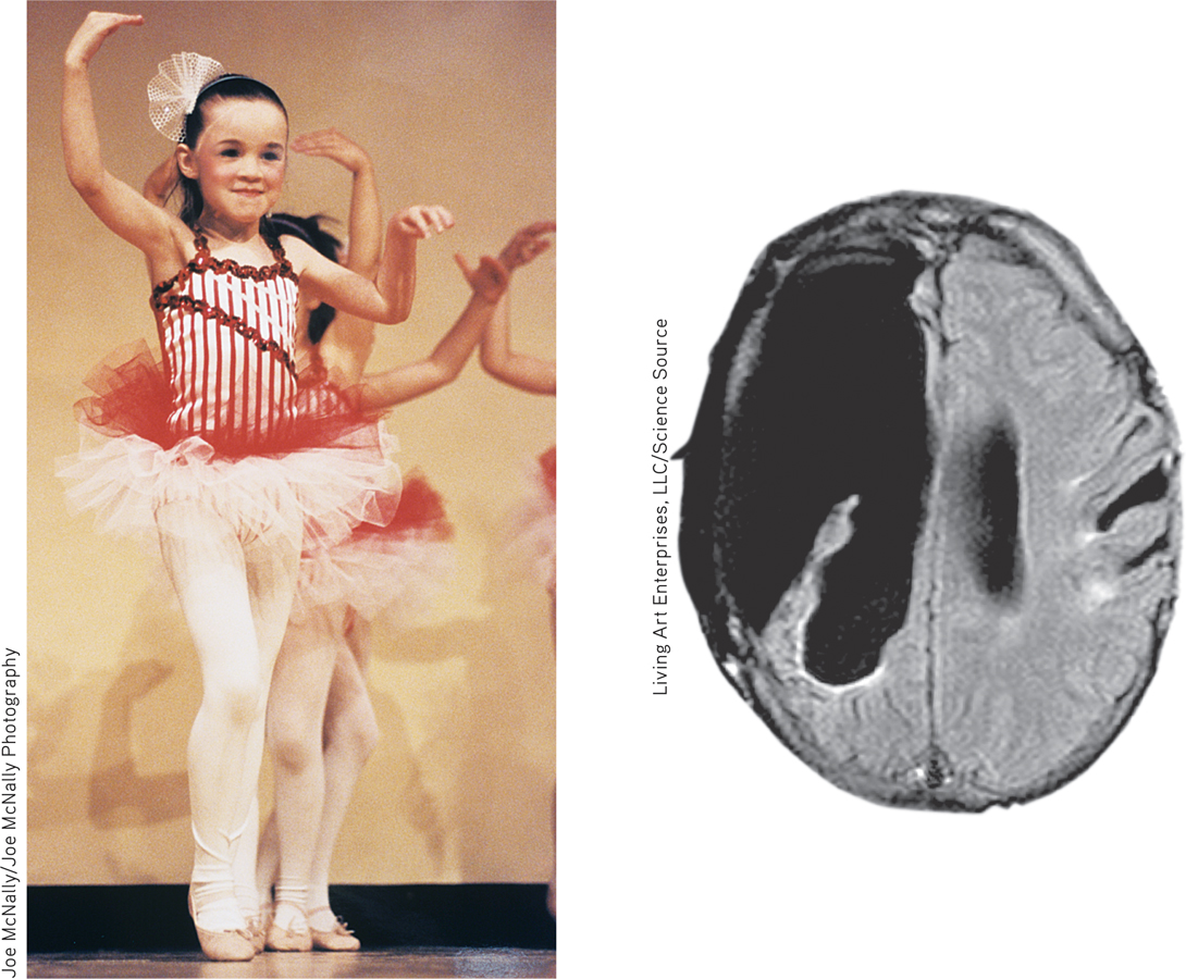
Brain plasticity This 6-
The brain’s plasticity is good news for those blind or deaf. Blindness or deafness makes their unused brain areas available for other uses (Amedi et al., 2005). If a blind person uses one finger to read Braille, the brain area dedicated to that finger expands as the sense of touch invades the visual cortex that normally helps people see (Barinaga, 1992a; Sadato et al., 1996). Plasticity also helps explain why some studies have found that deaf people have enhanced peripheral and motion-
82
Similar reassignment may occur when disease or damage frees up other brain areas normally dedicated to specific functions. If a slow-
neurogenesis the formation of new neurons.
Although the brain often attempts self-
Cold War nuclear tests between 1945 and 1963 oddly later enabled scientists to confirm the birth of new brain neurons. The blasts released radioactive carbon isotopes, which carbon-
Master stem cells that can develop into any type of brain cell have also been discovered in the human embryo. If mass-
Our Divided Brain
2-
Our brain’s look-
Splitting the Brain
In 1961, Los Angeles neurosurgeons Philip Vogel and Joseph Bogen speculated that major epileptic seizures were caused by an amplification of abnormal brain activity bouncing back and forth between the two cerebral hemispheres, which work together as a whole system. If so, they wondered, could they put an end to this biological tennis match by severing the corpus callosum, the wide band of axon fibers connecting the two hemispheres and carrying messages between them (see FIGURE 2.32)? Vogel and Bogen knew that psychologists Roger Sperry, Ronald Myers, and Michael Gazzaniga had divided cats’ and monkeys’ brains in this manner, with no serious ill effects.
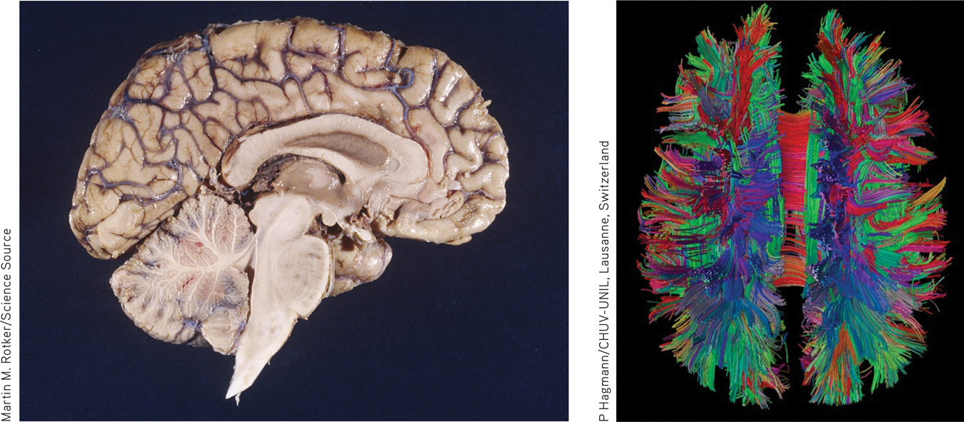
The corpus callosum This large band of neural fibers connects the two brain hemispheres. To photograph the half brain at left, a surgeon separated the hemispheres by cutting through the corpus callosum and lower brain regions. The high-
corpus callosum [KOR-pus kah-LOW-sum] the large band of neural fibers connecting the two brain hemispheres and carrying messages between them.
83
split brain a condition resulting from surgery that isolates the brain’s two hemispheres by cutting the fibers (mainly those of the corpus callosum) connecting them.
So the surgeons operated. The result? The seizures all but disappeared. The patients with these split brains were surprisingly normal, their personality and intellect hardly affected. Waking from surgery, one even joked that he had a “splitting headache” (Gazzaniga, 1967). By sharing their experiences, these patients have greatly expanded our understanding of interactions between the intact brain’s two hemispheres.
To appreciate these findings, we need to focus for a minute on the peculiar nature of our visual wiring, illustrated in FIGURE 2.33. Note that each eye receives sensory information from the entire visual field. But in each eye, information from the left half of your field of vision goes to your right hemisphere, and information from the right half of your visual field goes to your left hemisphere, which usually controls speech. Data received by either hemisphere are quickly transmitted to the other across the corpus callosum. In a person with a severed corpus callosum, this information-
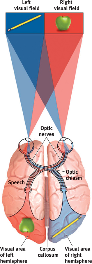
The information highway from eye to brain
Knowing these facts, Sperry and Gazzaniga could send information to a patient’s left or right hemisphere. As the person stared at a spot, they flashed a stimulus to its right or left. They could do this with you, too, but in your intact brain, the hemisphere receiving the information would instantly pass the news to the other side. Because the split-
In an early experiment, Gazzaniga (1967) asked these people to stare at a dot as he flashed HE•ART on a screen (FIGURE 2.34 below). Thus, HE appeared in their left visual field (which transmits to the right hemisphere) and ART in the right field (which transmits to the left hemisphere). When he then asked them to say what they had seen, the patients reported that they had seen ART. But when asked to point to the word they had seen, they were startled when their left hand (controlled by the right hemisphere) pointed to HE. Given an opportunity to express itself, each hemisphere indicated what it had seen. The right hemisphere (controlling the left hand) intuitively knew what it could not verbally report.
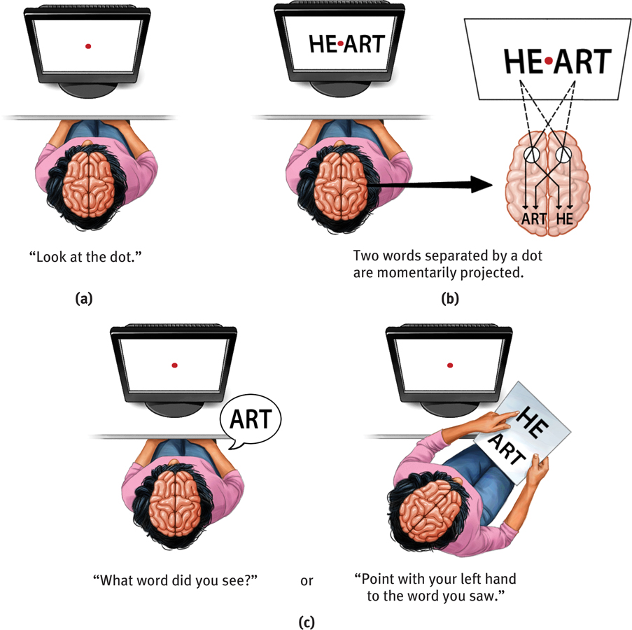
One skull, two minds When an experimenter flashes the word HEART across the visual field, a woman with a split brain verbally reports seeing the portion of the word transmitted to her left hemisphere. However, if asked to indicate with her left hand what she saw, she points to the portion of the word transmitted to her right hemisphere. (From Gazzaniga, 1983.)
84
“Do not let your left hand know what your right hand is doing.”
Matthew 6:3
When a picture of a spoon was flashed to their right hemisphere, the patients could not say what they had viewed. But when asked to identify what they had viewed by feeling an assortment of hidden objects with their left hand, they readily selected the spoon. If the experimenter said, “Correct!” the patient might reply, “What? Correct? How could I possibly pick out the correct object when I don’t know what I saw?” It is, of course, the left hemisphere doing the talking here, bewildered by what the nonverbal right hemisphere knows.
A few people who have had split-

Try this! People who have had split-
When the “two minds” are at odds, the left hemisphere does mental gymnastics to rationalize reactions it does not understand. If a patient follows an order (“Walk”) sent to the right hemisphere, a strange thing happens. The unaware left hemisphere doesn’t know why the patient begins walking. If asked, the patient doesn’t reply, “I don’t know.” Instead, the left hemisphere improvises—
85

 HOW WOULD YOU KNOW?Have you ever been asked if you are “left-brained” or “right-brained?” Consider this popular misconception with LaunchPad’s How Would You Know If People Can be “Left-Brained” or “Right-Brained”?
RETRIEVAL PRACTICE
- (1) If we flash a red light to the right hemisphere of a person with a split brain, and flash a green light to the left hemisphere, will each observe its own color? (2) Will the person be aware that the colors differ? (3) What will the person verbally report seeing?
1. yes, 2. no, 3. green
Right–
So, what about the 99.99+ percent of us with undivided brains? Does each of our hemispheres also perform distinct functions? Several different types of studies indicate they do. When a person performs a perceptual task, for example, brain waves, bloodflow, and glucose consumption reveal increased activity in the right hemisphere. When the person speaks or calculates, activity usually increases in the left hemisphere.
A dramatic demonstration of hemispheric specialization happens before some types of brain surgery. To locate the patient’s language centers, the surgeon injects a sedative into the neck artery feeding blood to the left hemisphere, which usually controls speech. Before the injection, the patient is lying down, arms in the air, chatting with the doctor. Can you predict what probably happens when the drug puts the left hemisphere to sleep? Within seconds, the person’s right arm falls limp. If the left hemisphere is controlling language, the patient will be speechless until the drug wears off. If the drug is injected into the artery to the right hemisphere, the left arm will fall limp, but the person will still be able to speak.
To the brain, language is language, whether spoken or signed. Just as hearing people usually use the left hemisphere to process spoken language, deaf people use the left hemisphere to process sign language (Corina et al., 1992; Hickok et al., 2001). Thus, a left hemisphere stroke disrupts a deaf person’s signing, much as it would disrupt a hearing person’s speaking (Corina, 1998).
Although the left hemisphere is adept at making quick, literal interpretations of language, the right hemisphere
- excels in making inferences (Beeman & Chiarello, 1998; Bowden & Beeman, 1998; Mason & Just, 2004). Primed with the flashed word foot, the left hemisphere will be especially quick to recognize the closely associated word heel. But if given an insight-like problem—“What word goes with boot, summer, and ground?”—the right hemisphere more quickly recognizes the solution: camp. As one patient explained after a right hemisphere stroke, “I understand words, but I’m missing the subtleties.” The right side of the brain is also better than the left at copying drawings, recognizing faces, noticing differences, perceiving emotion, and expressing emotion through the more expressive left side of the face. Right hemisphere damage can greatly disrupt these abilities.
- helps us modulate our speech to make meaning clear—as when we ask “What’s that in the road ahead?” instead of “What’s that in the road, a head?” (Heller, 1990).
- helps orchestrate our self-awareness. People who suffer partial paralysis will sometimes obstinately deny their impairment—strangely claiming they can move a paralyzed limb—if the damage is to the right hemisphere (Berti et al., 2005).
86
Simply looking at the two hemispheres, so alike to the naked eye, who would suppose they contribute uniquely to the harmony of the whole? Yet a variety of observations—
For a helpful animated review of this research, see LaunchPad’s PsychSim 6: Hemispheric Specialization.
THINKING CRITICALLY ABOUT
Handedness
2-13 What does research tell us about being left-handed? Is it advantageous to be right-handed?
Nearly 90 percent of us are primarily right-handed (Leask & Beaton, 2007; Medland et al., 2004; Peters et al., 2006). Most people also kick with their right foot and look through a microscope with their right eye. Some 10 percent of us (somewhat more among males, somewhat less among females) are left-handed. (A few people write with their right hand and throw a ball with their left, or vice versa.) Almost all right-handers (96 percent) process speech primarily in the left hemisphere, which tends to be the slightly larger hemisphere (Bishop, 2013). Left-handers are more diverse. Seven in ten process speech in the left hemisphere, as right-handers do. The rest either process language in the right hemisphere or use both hemispheres.
Is Handedness Inherited?
Judging from prehistoric human cave drawings, tools, and hand and arm bones, this veer to the right occurred long ago (Corballis, 1989; MacNeilage et al., 2009). Right-handedness prevails in all human cultures, and even in chimpanzees (Hopkins, 2013). Moreover, it appears prior to culture’s impact: More than 9 in 10 fetuses suck the right hand’s thumb (Hepper et al., 1990, 2004). Twin studies indicate only a small genetic influence on individual handedness (Vuoksimaa et al., 2009). But the universal prevalence of right-handers in humans and other primates suggests that either genes or some prenatal factors influence handedness.
So, Is It All Right to Be Left-Handed?
Judging by our everyday conversation, left-handedness is not all right. To be “coming out of left field” is hardly better than to be “gauche” (derived from the French word for “left”). On the other hand, right-handedness is “right on,” which any “righteous,” “right-hand man” “in his right mind” usually is.
Left-handers are more numerous than usual among those with reading disabilities, allergies, and migraine headaches (Geschwind & Behan, 1984). But in Iran, where students report which hand they write with when taking the university entrance exam, lefties have outperformed righties in all subjects (Noroozian et al., 2003). Left-handedness is also more common among musicians, mathematicians, professional baseball and cricket players, architects, and artists, including such luminaries as Michelangelo, Leonardo da Vinci, and Picasso.2 Although left-
RETRIEVAL PRACTICE
- Almost all right-handers process speech in the ______________ hemisphere; most left-handers process speech in the ______________ hemisphere.
left; left—
***
87
In this chapter we have glimpsed an overriding principle: Everything psychological is simultaneously biological. We have focused on how our thoughts, feelings, and actions arise from our specialized yet integrated brain. In chapters to come, we will further explore the significance of the biological revolution in psychology.
From nineteenth-
Much as gas and air can give rise to something different—
“All psychological phenomena are caused by the brain, but many are better understood at the level of the mind.”
Tweet from psychologist Steven Pinker, June 10, 2013
Cells cannot be fully explained by the actions of atoms, nor minds by the activity of cells. Psychology is rooted in biology, which is rooted in chemistry, which is rooted in physics. Yet psychology is more than applied physics. As Jerome Kagan (1998) reminded us, the meaning of the Gettysburg Address is not reducible to neural activity. Sexual love is more than blood flooding to the genitals. Morality and responsibility become possible when we understand the mind as a “holistic system,” said Sperry (1992) (FIGURE 2.36). We are not mere jabbering robots. Brains make thoughts. And thoughts change brains.
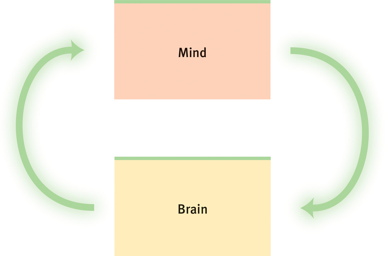
Mind and brain as holistic system In Roger Sperry’s view, the brain creates and controls the emergent mind, which in turn influences the brain. (Think vividly about biting into a lemon and you may salivate.)
“‘Was the cause psychological or biological?’ is the wrong question when assigning responsibility for an action. All psychological states are also biological ones.”
Psychologists John Monterosso and Barry Schwartz, “Did Your Brain Make You Do It?” 2012
The mind seeking to understand the brain—
88
REVIEW: The Cerebral Cortex and Our Divided Brain

|
REVIEW | The Cerebral Cortex and Our Divided Brain |
LEARNING OBJECTIVES
RETRIEVAL PRACTICE Take a moment to answer each of these Learning Objective Questions (repeated here from within this section). Then click the 'show answer' button to check your answers. Research suggests that trying to answer these questions on your own will improve your long-
2-
The cerebral cortex has two hemispheres, and each hemisphere has four lobes: the frontal, parietal, occipital, and temporal. Each lobe performs many functions and interacts with other areas of the cortex.
The motor cortex, at the rear of the frontal lobes, controls voluntary movements. The somatosensory cortex, at the front of the parietal lobes, registers and processes body touch and movement sensations. Body parts requiring precise control (in the motor cortex) or those that are especially sensitive (in the somatosensory cortex) occupy the greatest amount of space.
Most of the brain’s cortex—the major portion of each of the four lobes—is devoted to uncommitted association areas, which integrate information involved in learning, remembering, thinking, and other higher-level functions. Our mental experiences arise from coordinated brain activity.
2-
If one hemisphere is damaged early in life, the other will pick up many of its functions by reorganizing or building new pathways. This plasticity diminishes later in life. The brain sometimes mends itself by forming new neurons, a process known as neurogenesis.
2-
Split-brain research (experiments on people with a severed corpus callosum) has confirmed that in most people, the left hemisphere is the more verbal, and that the right hemisphere excels in visual perception and the recognition of emotion. Studies of healthy people with intact brains confirm that each hemisphere makes unique contributions to the integrated functioning of the brain.
2-
Some 10 percent of us (somewhat more among males, somewhat less among females) are left-handed. Handedness appears to be influenced by genetic or prenatal factors. Most left-handers process speech in the left hemisphere, as right-handers do, but some do so in the right hemisphere or use both hemispheres. Left-handers are more likely to be among those with reading disabilities, allergies, and migraine headaches, but sometimes do better academically. Left-handedness is also more common among musicians, mathematicians, architects, artists, and in professional baseball and cricket players. The pros and cons of being left-handed seem roughly equal.
TERMS AND CONCEPTS TO REMEMBER
RETRIEVAL PRACTICE Match each of the terms on the left with its definition on the right. Click on the term first and then click on the matching definition. As you match them correctly they will move to the bottom of the activity.
Question
cerebral [seh-REE-bruhl] cortex frontal lobes parietal [puh-RYE-uh-tuhl] lobes occipital [ahk-SIP-uh-tuhl] lobes temporal lobes motor cortex somatosensory cortex association areas plasticity neurogenesis corpus callosum [KOR-pus kah-LOW-sum] split brain | the brain’s ability to change, especially during childhood, by reorganizing after damage or by building new pathways based on experience. the large band of neural fibers connecting the two brain hemispheres and carrying messages between them. an area at the rear of the frontal lobes that controls voluntary movements. portion of the cerebral cortex lying just behind the forehead; involved in speaking and muscle movements and in making plans and judgments. areas of the cerebral cortex that are not involved in primary motor or sensory functions; rather, they are involved in higher mental functions such as learning, remembering, thinking, and speaking. portion of the cerebral cortex lying at the back of the head; includes areas that receive information from the visual fields. the intricate fabric of interconnected neural cells covering the cerebral hemispheres; the body’s ultimate control and information-processing center. a condition resulting from surgery that isolates the brain’s two hemispheres by cutting the fibers (mainly those of the corpus callosum) connecting them. portion of the cerebral cortex lying roughly above the ears; includes the auditory areas, each receiving information primarily from the opposite ear. area at the front of the parietal lobes that registers and processes body touch and movement sensations. the formation of new neurons. portion of the cerebral cortex lying at the top of the head and toward the rear; receives sensory input for touch and body position. |
Use  to create your personalized study plan, which will direct you to the resources that will help you most in
.
to create your personalized study plan, which will direct you to the resources that will help you most in
.
TEST
YOUR-
SELF THE BIOLOGY OF MIND
Test yourself repeatedly throughout your studies. This will not only help you figure out what you know and don’t know; the testing itself will help you learn and remember the information more effectively thanks to the testing effect.
 Neural and Hormonal Systems
Question
1. The neuron fiber that passes messages through its branches to other neurons or to muscles and glands is the .
Question
2. The tiny space between the axon of one neuron and the dendrite or cell body of another is called the
| A. |
| B. |
| C. |
| D. |
Question
3. Regarding a neuron’s response to stimulation, the intensity of the stimulus determines
| A. |
| B. |
| C. |
| D. |
Question
4. In a sending neuron, when an action potential reaches an axon terminal, the impulse triggers the release of chemical messengers called .
Question
5. Endorphins are released in the brain in response to
| A. |
| B. |
| C. |
| D. |
Question
6. The autonomic nervous system controls internal functions, such as heart rate and glandular activity. The word autonomic means
| A. |
| B. |
| C. |
| D. |
Question
7. The sympathetic nervous system arouses us for action and the parasympathetic nervous system calms us down. Together, the two systems make up the nervous system.
Question
8. The neurons of the spinal cord are part of the nervous system.
Question
9. The most influential endocrine gland, known as the master gland, is the
| A. |
| B. |
| C. |
| D. |
Question
10. The secrete(s) epinephrine and norepinephrine, helping to arouse the body during times of stress.
Tools of Discovery and Older Brain Structures
Question
11. The part of the brainstem that controls heartbeat and breathing is the
| A. |
| B. |
| C. |
| D. |
Question
12. The thalamus functions as a
| A. |
| B. |
| C. |
| D. |
Question
13. The lower brain structure that governs arousal is the
| A. |
| B. |
| C. |
| D. |
Question
14. The part of the brain that coordinates voluntary movement and enables nonverbal learning and memory is the .
Question
15. Two parts of the limbic system are the amygdala and the
| A. |
| B. |
| C. |
| D. |
Question
16. A cat’s ferocious response to electrical brain stimulation would lead you to suppose the electrode had touched the .
Question
17. The neural structure that most directly regulates eating, drinking, and body temperature is the
| A. |
| B. |
| C. |
| D. |
Question
18. The initial reward center discovered by Olds and Milner was located in the .
The Cerebral Cortex and Our Divided Brain
Question
19. If a neurosurgeon stimulated your right motor cortex, you would most likely
| A. |
| B. |
| C. |
| D. |
Question
20. How do different neural networks communicate with one another to let you respond when a friend greets you at a party?
The visual cortex is a neural network of sensory neurons connected via interneurons to other neural networks, including auditory networks. This allows you to integrate visual and auditory information to respond when a friend you recognize greets you at a party.
Question
21. Which of the following body regions has the greatest representation in the somatosensory cortex?
| A. |
| B. |
| C. |
| D. |
Question
22. Judging and planning are enabled by the lobes.
Question
23. What would it be like to talk on the phone if you didn’t have temporal lobe association areas? What would you hear? What would you understand?
You would hear sounds, but without the temporal lobe association areas you would be unable to make sense of what you were hearing.
Question
24. The “uncommitted” areas that make up about three-fourths of the cerebral cortex are called .
Question
25. Plasticity is especially evident in the brains of
| A. |
| B. |
| C. |
| D. |
Question
26. An experimenter flashes the word HERON across the visual field of a man whose corpus callosum has been severed. HER is transmitted to his right hemisphere and ON to his left hemisphere. When asked to indicate what he saw, the man says he saw but points to .
Question
27. Studies of people with split brains and brain scans of those with undivided brains indicate that the left hemisphere excels in
| A. |
| B. |
| C. |
| D. |
Question
28. Damage to the brain’s right hemisphere is most likely to reduce a person’s ability to
| A. |
| B. |
| C. |
| D. |