19.2 Information Processing in the Eye and Brain
Please continue to the next section.
Retinal Processing
19-
Imagine that you could follow behind a single light-
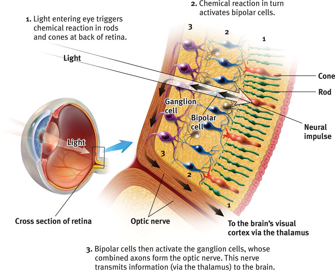
The retina’s reaction to light

The blind spot
RETRIEVAL PRACTICE
- There are no receptor cells where the optic nerve leaves the eye. This creates a blind spot in your vision. To demonstrate, first close your left eye, look at the spot above, and move your face away to a distance at which one of the cars disappears. (Which one do you predict it will be?) Repeat with your right eye closed—and note that now the other car disappears. Can you explain why?
Your blind spot is on the nose side of each retina, which means that objects to your right may fall onto the right eye’s blind spot. Objects to your left may fall on the left eye’s blind spot. The blind spot does not normally impair your vision, because your eyes are moving and because one eye catches what the other misses.
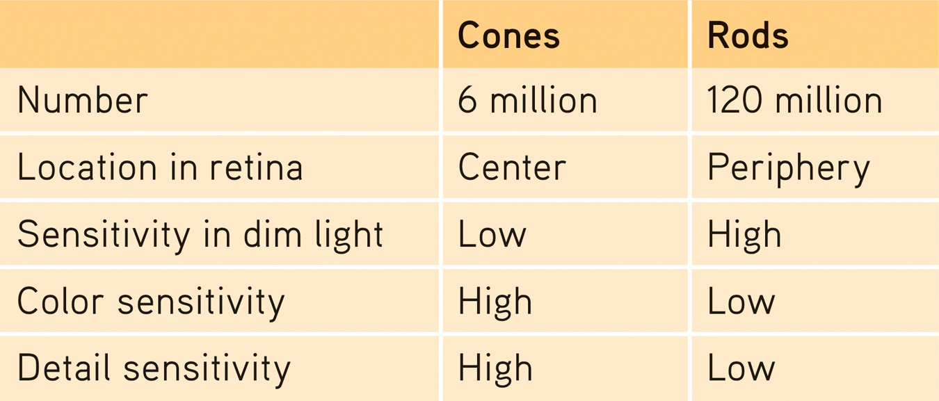
Rods and cones differ in where they’re found and in what they do (TABLE 19.1). Cones cluster in and around the fovea, the retina’s area of central focus (see Figure 19.3). Many cones have their own hotline to the brain: Each cone transmits its message to a single bipolar cell. That cell helps relay the cone’s individual message to the visual cortex, which devotes a large area to input from the fovea. These direct connections preserve the cones’ precise information, making them better able to detect fine detail.
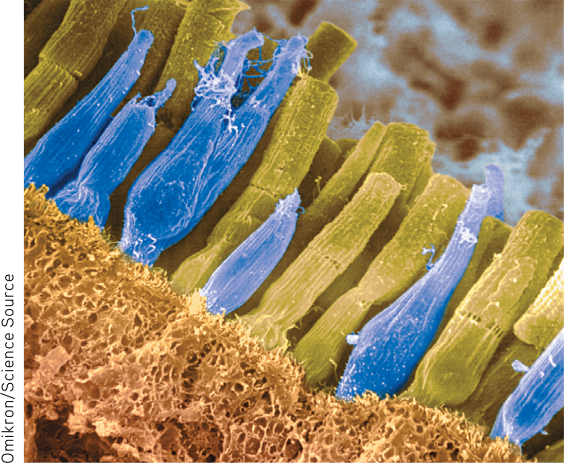
Receptors in the Human Eye: Rod-
Rods don’t have dedicated hotlines. Rods share bipolar cells which send combined messages. To experience this rod-
Cones also enable you to perceive color. In dim light they become ineffectual, so you see no colors. Rods, which enable black-
When you enter a darkened theater or turn off the light at night, your eyes adapt. Your pupils dilate to allow more light to reach your retina, but it typically takes 20 minutes or more before your eyes fully adapt. You can demonstrate dark adaptation by closing or covering one eye for up to 20 minutes. Then make the light in the room not quite bright enough to read this book with your open eye. Now open the dark-
To summarize: The retina’s neural layers don’t just pass along electrical impulses. They also help to encode and analyze sensory information. (The third neural layer in a frog’s eye, for example, contains the “bug detector” cells that fire only in response to moving fly-
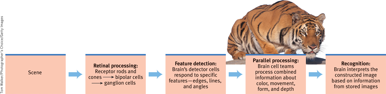
- After processing by your retina’s nearly 130 million receptor rods and cones, information travels forward again, to your bipolar cells.
- From there, it moves to your eye’s million or so ganglion cells, and through their axons making up the optic nerve to your brain.
- After a momentary stop-off in the thalamus, the information travels to your visual cortex. Any given retinal area relays its information to a corresponding location in your visual cortex, in the occipital lobe at the back of your brain (FIGURE 19.6).
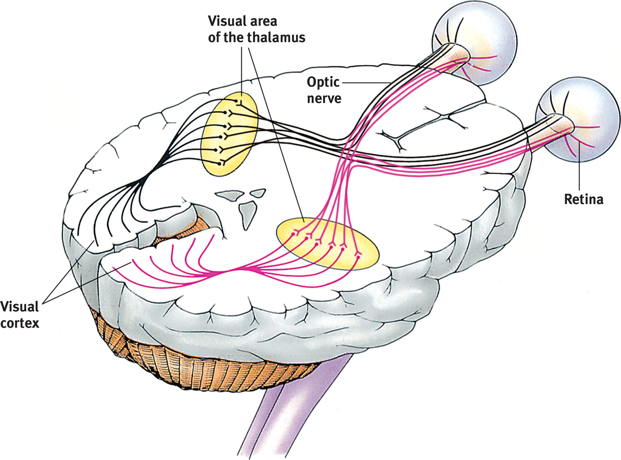
 Figure 19.6
Pathway from the eyes to the visual cortex Ganglion axons forming the optic nerve run to the thalamus, where they synapse with neurons that run to the visual cortex.

The same sensitivity that enables retinal cells to fire messages can lead them to misfire, as you can demonstrate. Turn your eyes to the left, close them, and then gently rub the right side of your right eyelid with your fingertip. Note the patch of light to the left, moving as your finger moves.
Why do you see light? Why at the left? This happens because your retinal cells are so responsive that even pressure triggers them. But your brain interprets their firing as light. Moreover, it interprets the light as coming from the left—
RETRIEVAL PRACTICE
- Some nocturnal animals, such as toads, mice, rats, and bats, have impressive night vision thanks to having many more ____________ (rods/cones) than ____________ (rods/cones) in their retinas. These creatures probably have very poor ____________ (color/black-and-white) vision.
rods; cones; color
- Cats are able to open their ____________ much wider than we can, which allows more light into their eyes so they can see better at night.
pupils
Color Processing
19-
One of vision’s most basic and intriguing mysteries is how we see the world in color. In everyday conversation, we talk as though objects possess color: “A tomato is red.” Recall the old question, “If a tree falls in the forest and no one hears it, does it make a sound?” We can ask the same of color: If no one sees the tomato, is it red?
The answer is No. First, the tomato is everything but red, because it rejects (reflects) the long wavelengths of red. Second, the tomato’s color is our mental construction. As Isaac Newton (1704) noted, “The [light] rays are not colored.” Like all aspects of vision, our perception of color resides not in the object itself but in the theater of our brains, as evidenced by our dreaming in color.
“Only mind has sight and hearing; all things else are deaf and blind.”
Epicharmus, Fragments, 550 b.c.e.
How, from the light energy striking the retina, does our brain construct our experience of color—
Modern detective work on the mystery of color vision began in the nineteenth century, when Hermann von Helmholtz built on the insights of an English physicist, Thomas Young. Any color can be created by combinations of different amounts of light waves of three primary colors—
Most people with color-
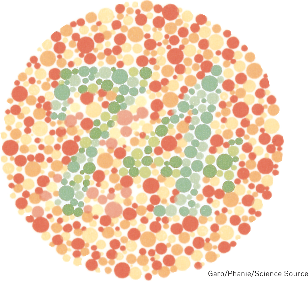
Color-
But why do people blind to red and green often still see yellow? And why does yellow appear to be a pure color and not a mixture of red and green, the way purple is of red and blue? Trichromatic theory left some parts of the color vision mystery unsolved, and this sparked researcher Ewald Hering’s curiosity.
Hering, a physiologist, found a clue in afterimages. Stare at a green square for a while and then look at a white sheet of paper, and you will see red, green’s opponent color. Stare at a yellow square and its opponent color, blue, will appear on the white paper. (To experience this, try the flag demonstration in FIGURE 19.8.) Hering formed another hypothesis: There must be two additional color processes, one responsible for red-

Afterimage effect Stare at the center of the flag for a minute and then shift your eyes to the dot in the white space beside it. What do you see? (After tiring your neural response to black, green, and yellow, you should see their opponent colors.) Stare at a white wall and note how the size of the flag grows with the projection distance.
Indeed, a century later, researchers also confirmed Hering’s opponent-process theory. Three sets of opponent retinal processes—
So how do we explain afterimages, such as in the flag demonstration? By staring at green, we tire our green response. When we then stare at white (which contains all colors, including red), only the red part of the green-
The present solution to the mystery of color vision is therefore roughly this: Color processing occurs in two stages.
- The retina’s red, green, and blue cones respond in varying degrees to different color stimuli, as the Young-Helmholtz trichromatic theory suggested.
- The cones’ responses are then processed by opponent-process cells, as Hering’s theory proposed.
 For an interactive review and demonstration of these color vision principles, visit LaunchPad’s PsychSim 6: Colorful World.
RETRIEVAL PRACTICE
- What are two key theories of color vision? Are they contradictory or complementary? Explain.
The Young-
Feature Detection
19-
Once upon a time, scientists believed that the brain was like a movie screen, on which the eye projected images. But then along came David Hubel and Torsten Wiesel (1979), who showed that our brain’s computing system deconstructs visual images and then reassembles them. Hubel and Wiesel received a Nobel Prize for their work on feature detectors, nerve cells in the brain that respond to a scene’s specific features—
Using microelectrodes, they had discovered that some neurons fired actively when cats were shown lines at one angle, while other neurons responded to lines at a different angle. They surmised that these specialized neurons in the occipital lobe’s visual cortex—
One temporal lobe area by your right ear (FIGURE 19.9) enables you to perceive faces and, thanks to a specialized neural network, to recognize them from varied viewpoints (Connor, 2010). If stimulated in this area, you might spontaneously see faces. If this region were damaged, you might recognize other forms and objects, but not familiar faces.
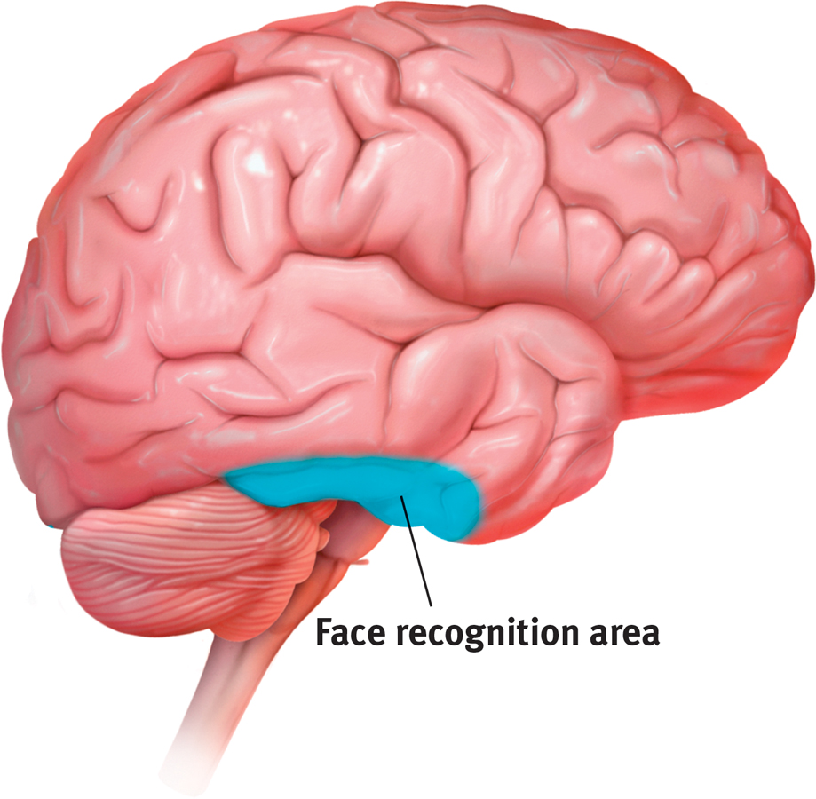
Face recognition processing In social animals such as humans, a large right temporal lobe area (shown here in a right-
Researchers can temporarily disrupt the brain’s face-
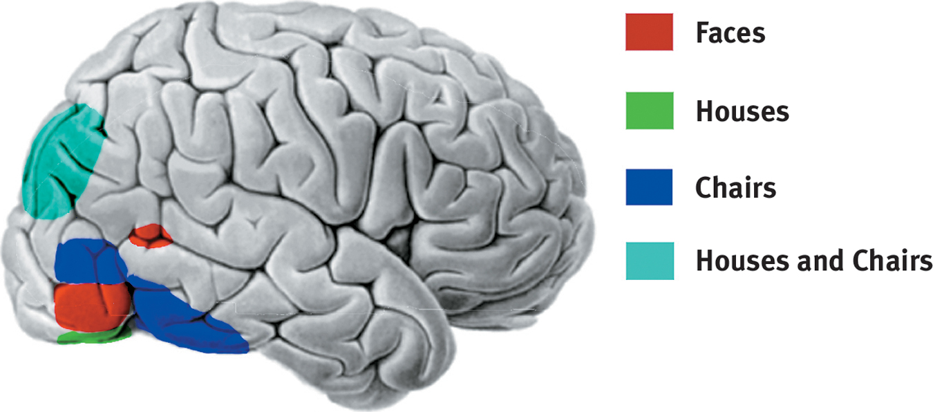
The telltale brain Looking at faces, houses, and chairs activates different brain areas in this right-
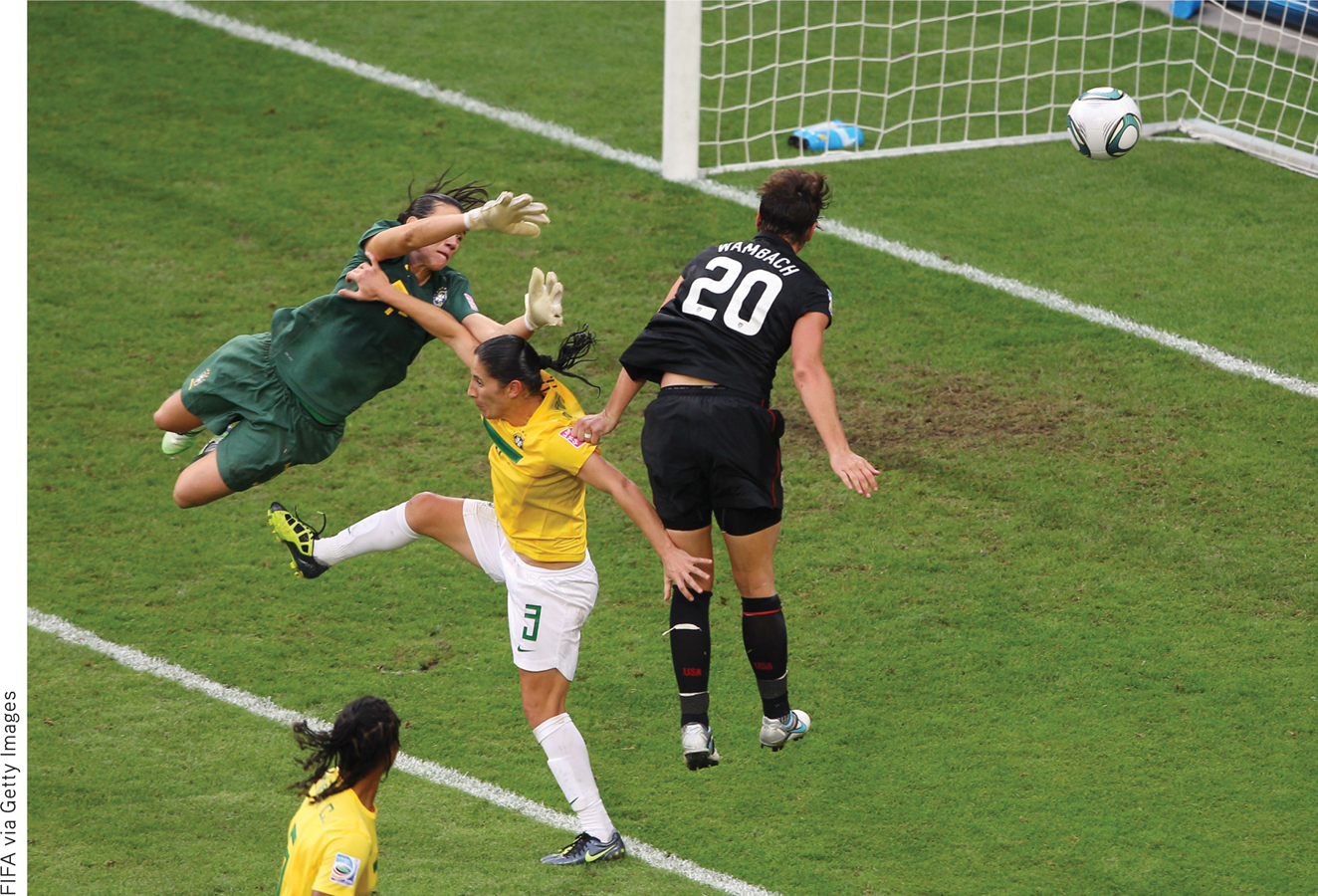
For biologically important objects and events, monkey brains (and surely ours as well) have a “vast visual encyclopedia” distributed as specialized cells (Perrett et al., 1988, 1992, 1994). These cells respond to one type of stimulus, such as a specific gaze, head angle, posture, or body movement. Other supercell clusters integrate this information and fire only when the cues collectively indicate the direction of someone’s attention and approach. This instant analysis, which aided our ancestors’ survival, also helps a soccer player anticipate where to strike the ball, and a driver anticipate a pedestrian’s next movement.
Parallel Processing
19-
Our brain achieves these and other remarkable feats by parallel processing: doing many things at once. To analyze a visual scene, the brain divides it into subdimensions—
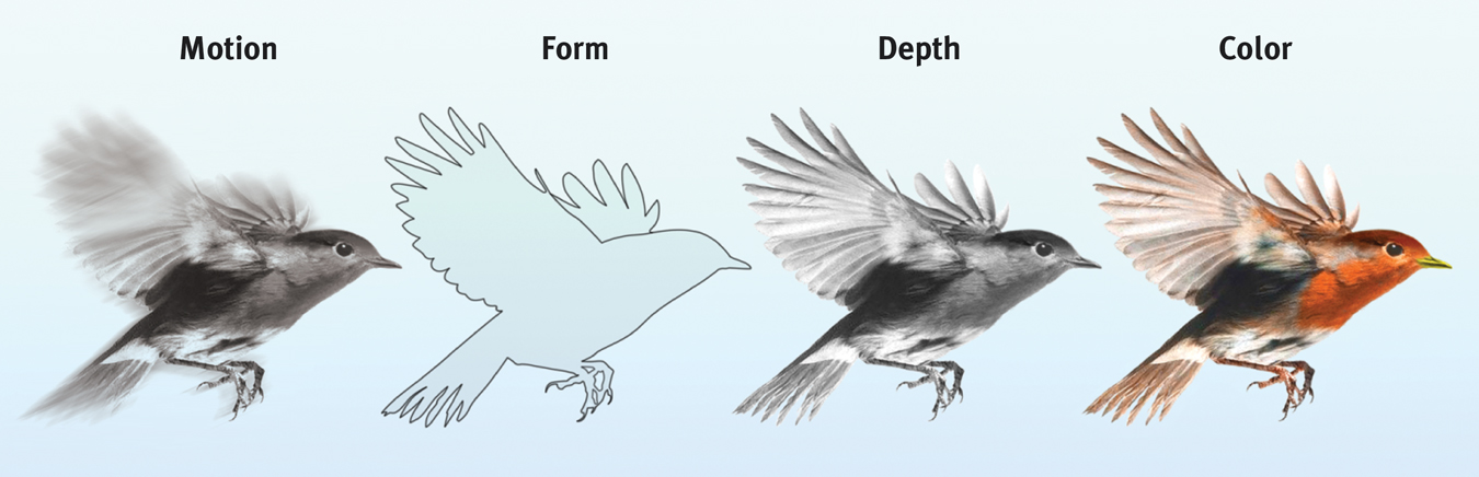
Parallel processing Studies of patients with brain damage suggest that the brain delegates the work of processing motion, form, depth, and color to different areas. After taking a scene apart, the brain integrates these subdimensions into the perceived image. How does the brain do this? The answer to this question is the Holy Grail of vision research.
To recognize a face, your brain integrates information projected by your retinas to several visual cortex areas, compares it to stored information, and enables you to recognize the face: Grandmother! Scientists have debated whether this stored information is contained in a single cell or, more likely, distributed over a vast network of cells. Some supercells—
Destroy or disable a neural workstation for a visual subtask, and something peculiar results, as happened to “Mrs. M.” (Hoffman, 1998). Since a stroke damaged areas near the rear of both sides of her brain, she has been unable to perceive movement. People in a room seem “suddenly here or there but I have not seen them moving.” Pouring tea into a cup is a challenge because the fluid appears frozen—
After stroke or surgery has damaged the brain’s visual cortex, others have experienced blindsight. Shown a series of sticks, they report seeing nothing. Yet when asked to guess whether the sticks are vertical or horizontal, their visual intuition typically offers the correct response. When told, “You got them all right,” they are astounded. There is, it seems, a second “mind”—a parallel processing system—
For a 4-minute depiction of a blindsight patient, visit the Launch-Pad Video—Blindsight: Seeing Without Awareness.
***
“I am … wonderfully made.”
King David, Psalm 139:14
Think about the wonders of visual processing. As you read this page, the letters are transmitted by reflected light rays onto your retina, which triggers a process that sends formless nerve impulses to several areas of your brain, which integrates the information and decodes meaning, thus completing the transfer of information across time and space from my mind to your mind (FIGURE 19.12). That all of this happens instantly, effortlessly, and continuously is indeed awesome. As Roger Sperry (1985) observed, the “insights of science give added, not lessened, reasons for awe, respect, and reverence.”
A simplified summary of visual information processing
RETRIEVAL PRACTICE
- What is the rapid sequence of events that occurs when you see and recognize a friend?
Light waves reflect off the person and travel into your eye, where the receptor cells in your retina convert the light waves’ energy into neural impulses sent to your brain. Your brain processes the subdimensions of this visual input—