25.1 Memory Storage
25-
In Arthur Conan Doyle’s A Study in Scarlet, Sherlock Holmes offers a popular theory of memory capacity:
I consider that a man’s brain originally is like a little empty attic, and you have to stock it with such furniture as you choose…. It is a mistake to think that that little room has elastic walls and can distend to any extent. Depend upon it, there comes a time when for every addition of knowledge you forget something that you knew before.
Contrary to Holmes’ “memory model,” our capacity for storing long-
Retaining Information in the Brain
I [DM] marveled at my aging mother-
For a time, some surgeons and memory researchers recorded patients’ seemingly vivid memories triggered by brain stimulation during surgery. Did this prove that our whole past, not just well-
“Our memories are flexible and super-imposable, a panoramic blackboard with an endless supply of chalk and erasers.”
Elizabeth Loftus and Katherine Ketcham, The Myth of Repressed Memory, 1994
The point to remember: Despite the brain’s vast storage capacity, we do not store information as libraries store their books, in single, precise locations. Instead, brain networks encode, store, and retrieve the information that forms our complex memories.
Explicit Memory System: The Frontal Lobes and Hippocampus
25-
The network that processes and stores your explicit memories for facts and episodes includes your frontal lobes and hippocampus. When you summon up a mental encore of a past experience, many brain regions send input to your frontal lobes for working memory processing (Fink et al., 1996; Gabrieli et al., 1996; Markowitsch, 1995). The left and right frontal lobes process different types of memories. Recalling a password and holding it in working memory, for example, would activate the left frontal lobe. Calling up a visual party scene would more likely activate the right frontal lobe.
Cognitive neuroscientists have found that the hippocampus, a temporal-
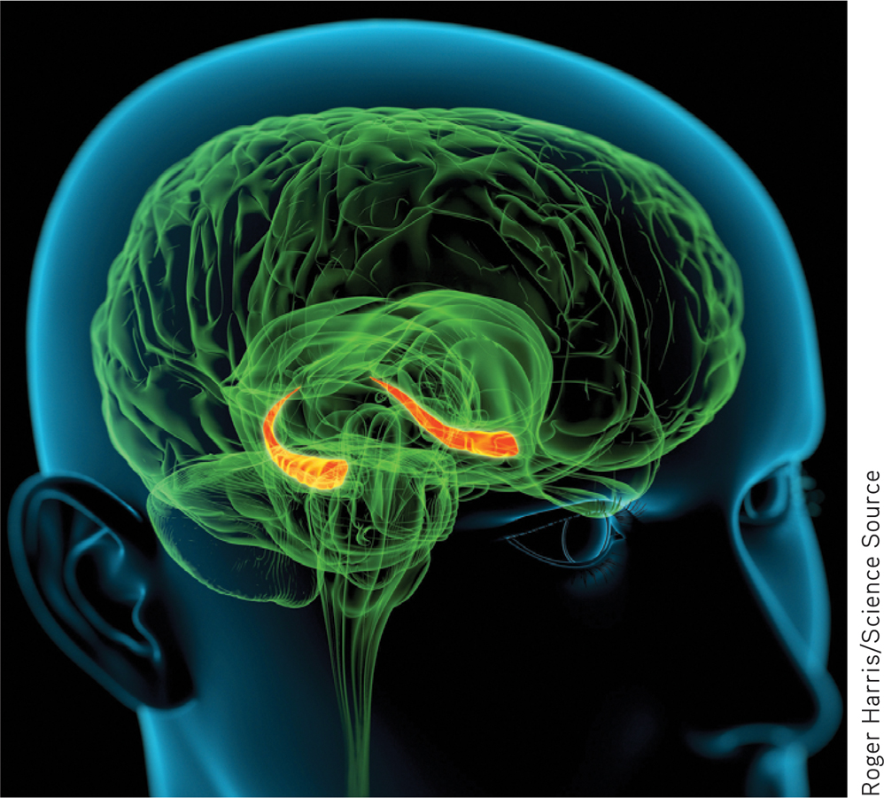
 Figure 25.1
The hippocampus Explicit memories for facts and episodes are processed in the hippocampus (orange structure) and fed to other brain regions for storage.

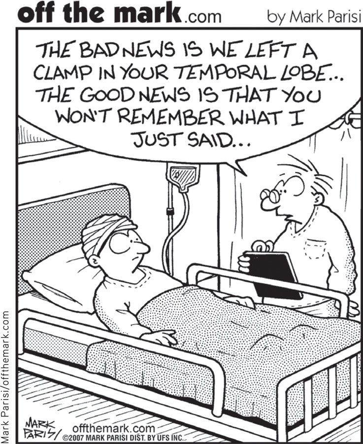
Damage to this structure therefore disrupts recall of explicit memories. Chickadees and other birds can store food in hundreds of places and return to these unmarked caches months later—
Subregions of the hippocampus also serve different functions. One part is active as people learn to associate names with faces (Zeineh et al., 2003). Another part is active as memory champions engage in spatial mnemonics (Maguire et al., 2003b). The rear area, which processes spatial memory, grows bigger the longer a London cabbie has navigated the maze of streets (Woolett & Maguire, 2011).
Memories are not permanently stored in the hippocampus. Instead, this structure seems to act as a loading dock where the brain registers and temporarily holds the elements of a remembered episode—
Sleep supports memory consolidation. During deep sleep, the hippocampus processes memories for later retrieval. After a training experience, the greater the hippocampus activity during sleep, the better the next day’s memory will be (Peigneux et al., 2004). Researchers have watched the hippocampus and brain cortex displaying simultaneous activity rhythms during sleep, as if they were having a dialogue (Euston et al., 2007; Mehta, 2007). They suspect that the brain is replaying the day’s experiences as it transfers them to the cortex for long-
Implicit Memory System: The Cerebellum and Basal Ganglia
25-
Your hippocampus and frontal lobes are processing sites for your explicit memories. But you could lose those areas and still, thanks to automatic (not effortful) processing, lay down implicit memories for skills and newly conditioned associations. Joseph LeDoux (1996) recounted the story of a brain-
The cerebellum plays a key role in forming and storing the implicit memories created by classical conditioning. With a damaged cerebellum, people cannot develop certain conditioned reflexes, such as associating a tone with an impending puff of air—
The basal ganglia, deep brain structures involved in motor movement, facilitate formation of our procedural memories for skills (Mishkin, 1982; Mishkin et al., 1997). The basal ganglia receive input from the cortex but do not return the favor of sending information back to the cortex for conscious awareness of procedural learning. If you have learned how to ride a bike, thank your basal ganglia.
Our implicit memory system, enabled by the cerebellum and basal ganglia, helps explain why the reactions and skills we learned during infancy reach far into our future. Yet as adults, our conscious memory of our first three years is blank, an experience called infantile amnesia. In one study, events children experienced and discussed with their mothers at age 3 were 60 percent remembered at age 7 but only 34 percent remembered at age 9 (Bauer et al., 2007). Two influences contribute to infantile amnesia: First, we index much of our explicit memory using words that nonspeaking children have not learned. Second, the hippocampus is one of the last brain structures to mature, and as it does, more gets retained (Akers et al., 2014).
RETRIEVAL PRACTICE
- Which parts of the brain are important for implicit memory processing, and which parts play a key role in explicit memory processing?
The cerebellum and basal ganglia are important for implicit memory processing and the frontal lobes and hippocampus are key to explicit memory formation.
- Your friend has experienced brain damage in an accident. He can remember how to tie his shoes but has a hard time remembering anything told to him during a conversation. What’s going on here?
Our explicit conscious memories of facts and episodes differ from our implicit memories of skills (such as shoe tying) and classically conditioned responses. Our implicit memories are processed by more ancient brain areas, which apparently escaped damage during the accident.
The Amygdala, Emotions, and Memory
25-
Our emotions trigger stress hormones that influence memory formation. When we are excited or stressed, these hormones make more glucose energy available to fuel brain activity, signaling the brain that something important has happened. Moreover, stress hormones focus memory. Stress provokes the amygdala (two limbic system, emotion-
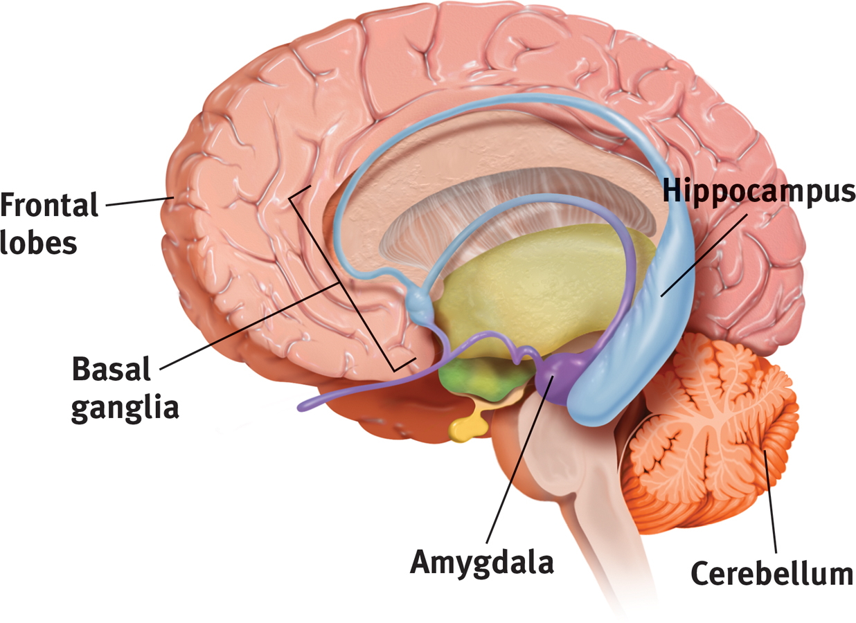
 Figure 25.2
Review key memory structures in the brain
Frontal lobes and hippocampus: explicit memory formation
Cerebellum and basal ganglia: implicit memory formation
Amygdala: emotion-
Emotions often persist without our conscious awareness of what caused them. In one ingenious experiment, patients with hippocampal damage (which left them unable to form new explicit memories) watched a sad film and later a happy film. After the viewing, they did not consciously recall the films, but the sad or happy emotion persisted (Feinstein et al., 2010).
Significantly stressful events can form almost indelible memories. After traumatic experiences—
Emotion-
The people who experienced a 1989 San Francisco earthquake did just that. A year and a half later, they had perfect recall of where they had been and what they were doing (verified by their recorded thoughts within a day or two of the quake). Others’ memories for the circumstances under which they merely heard about the quake were more prone to errors (Neisser et al., 1991; Palmer et al., 1991).
Which is more important—your experiences or your memories of them?
Our flashbulb memories are noteworthy for their vividness and our confidence in them. But as we relive, rehearse, and discuss them, these memories may come to err. With time, some errors crept into people’s 9/11 recollections (compared with their earlier reports taken right after 9/11). Mostly, however, people’s memories of 9/11 remained consistent over the next two to three years (Conway et al., 2009; Hirst et al., 2009; Kvavilashvili et al., 2009).
Dramatic experiences remain bright and clear in our memory in part because we rehearse them. We think about them and describe them to others. Memories of our best experiences, which we enjoy recalling and recounting, also endure (Storm & Jobe, 2012; Talarico & Moore, 2012). One study invited 1563 Boston Red Sox and New York Yankees fans to recall the baseball championship games between their two teams in 2003 (Yankees won) and 2004 (Red Sox won). Fans recalled much better the game their team won (Breslin & Safer, 2011).
Synaptic Changes
25-
As you read this module and think and learn about memory, your brain is changing. Given increased activity in particular pathways, neural interconnections are forming and strengthening.
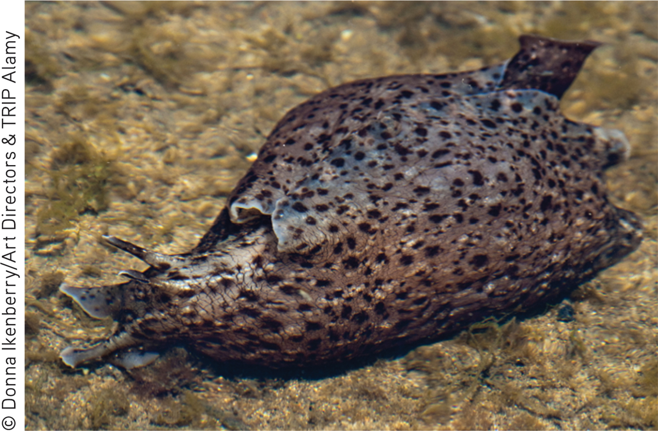
The quest to understand the physical basis of memory—
In experiments with people, rapidly stimulating certain memory-
- Drugs that block LTP interfere with learning (Lynch & Staubli, 1991).
- Mutant mice engineered to lack an enzyme needed for LTP couldn’t learn their way out of a maze (Silva et al., 1992).
- Rats given a drug that enhanced LTP learned a maze with half the usual number of mistakes (Service, 1994).
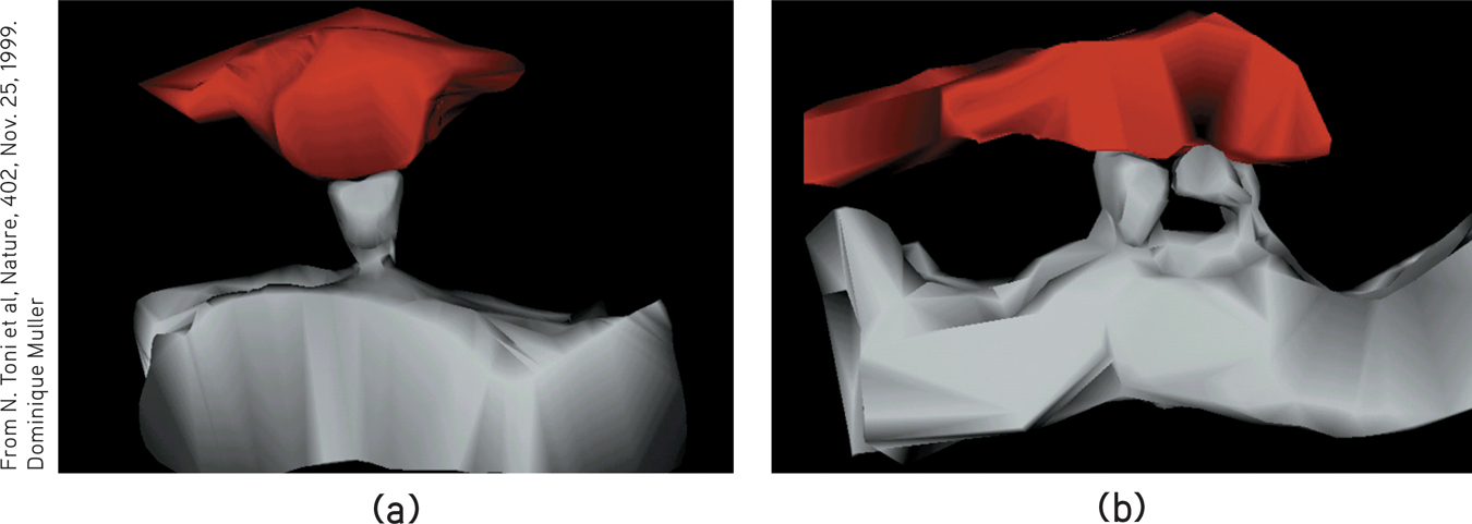
 Figure 25.3
Doubled receptor sites An electron microscope image (a) shows just one receptor site (gray) reaching toward a sending neuron before long-
After long-
Recently, I [DM] did a little test of memory consolidation. While on an operating table for a basketball-
Some memory-
One approach to improving memory focuses on drugs that boost the LTP-
Other people wish for memory-
In your lifetime, will you have access to safe and legal drugs that boost your fading memory without nasty side effects and without cluttering your mind with trivia best forgotten? That question has yet to be answered. But in the meantime, one effective, safe, and free memory enhancer is already available on your college campus: effective study techniques followed by adequate sleep!
FIGURE 25.4 summarizes the brain’s two-
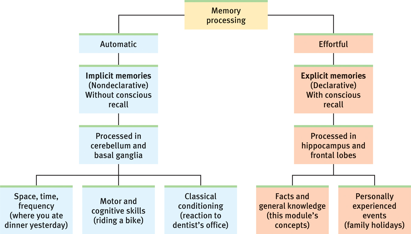
 Figure 25.4
Our two memory systems
RETRIEVAL PRACTICE
- Which brain area responds to stress hormones by helping to create stronger memories?
the amygdala
- The neural basis for learning and memory, found at the synapses in the brain’s memory-circuit connections, results from brief, rapid stimulation. It is called _________-_________ _________.
long-