true
Begin
true
Begin
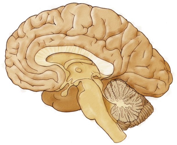
brainstem
Central structure of the brain (including the hindbrain, midbrain, thalamus, and hypothalamus) that is responsible for most life-sustaining, unconscious behavior
hindbrain
Evolutionarily the oldest part of the brain; contains the pons, medulla, reticular formation, and cerebellum, the structures that coordinate and control most voluntary and involuntary movements.
midbrain
Central part of the brain; contains neural circuits for hearing and seeing as well as for orienting movements.
afferent
Conducting toward a CNS structure.
efferent
Conducting away from a CNS structure.
pons
This structure, immediately rostral to the medulla, forms a physical bridge between the cerebellum and the rest of the brain. The pons regulates various autonomic functions, receives sensory information from the head and neck, controls various involuntary movements, and plays an important role in the regulation of sleep.
diencephalon
The between brain, which integrates sensory and motor information on its way to the cerebral cortex.
tectum
Roof (area above the ventricle) of the midbrain; its functions are sensory processing, particularly visual and auditory, and the production of orienting movements.
tegmentum
Floor (area below the ventricle) of the midbrain; a collection of nuclei with movement-related, species-specific, and pain perception functions.
orienting movement
Movement related to sensory inputs, such as turning the head to see the source of a sound.
cerebral cortex
Heavily folded and layered tissue that is the outer structure of the forebrain; composed of neocortex and allocortex.
thalamus
Diencephalon structure through which information from all sensory systems is organized, integrated, and projected into the appropriate region of the neocortex.
hypothalamus
Diencephalon structure that contains many nuclei associated with temperature regulation, eating, drinking, and sexual behavior.
dorsal
toward the back
superior
above
inferior
below
nuclei
A group of neurons forming a cluster that can be identified using special stains
forebrain
Evolutionarily the most recent addition to the brain; coordinates advanced cognitive functions such as thinking, planning, and language; contains the allocortex, neocortex, and basal ganglia.
allocortex
Part of the cerebral cortex (“outer bark”), composed of three or four layers; plays a role in controlling motivational and emotional states as well as in certain forms of memory.
neocortex
Most recently expanded outer layer (“new bark”) of the forebrain, composed of about six layers of gray matter. Its name is a misnomer, as it actually isn’t newer because it arose at the same time during evolution as other forms of the cortex. It is also called isocortex because it is almost always six-layered, with few exceptions.
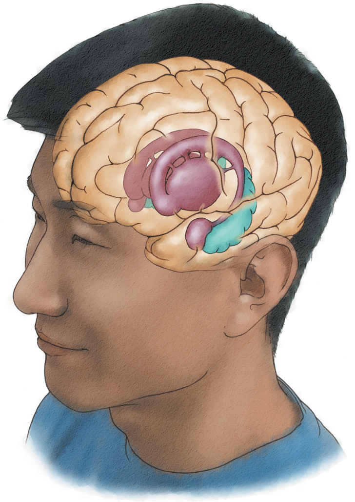
basal ganglia
Subcortical forebrain nuclei that coordinate voluntary movements of the limbs and body; connected to the thalamus and to the midbrain.
Parkinson disease
Disorder of the motor system correlated with a loss of dopamine from the substantia nigra and characterized by tremors, muscular rigidity, and a reduction in voluntary movement.
Dopamine
Amine neurotransmitter involved in coordinating movement, attention, learning, and reinforcing behaviors
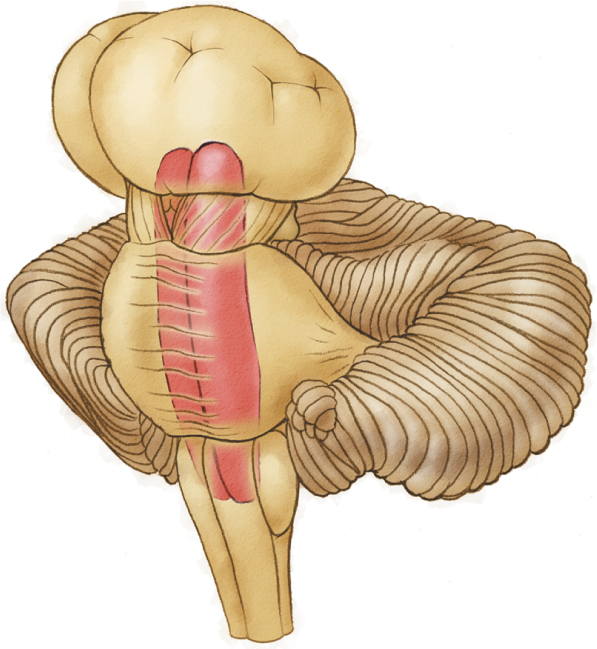
Periaqueductal gray matter
Nuclei in the midbrain that surround the cerebral aqueduct joining the third and fourth ventricles; PAG neurons contain circuits for species-typical behaviors (e.g., female sexual behavior) and play an important role in the modulation of pain.
red nucleus
The red nucleus plays a role in coordinating movement of the arms and hands.
substantia nigra
The substantia nigra connects to the basal ganglia and is critical for the initiation of voluntary motor movement. Parkinson disease is a movement disorder caused by the deterioration of dopamine cells in this area of the tegmentum.
periaqueductal gray matter
This distinct collection of cells that surrounds the cerebral aqueduct plays a role in both species-typical behaviors, such as female sexual behavior, and the modulation of pain.
reticular formation
This diffuse collection of cells that extend from the medulla (caudally) to the midbrain (rostrally) is involved in arousal, attention, and wakefulness.
Anatomy of the Brain
By: