Chapter 1. Finding Your Way Around the Brain
Finding Your Way Around the Brain
By:
You are about to embark on a journey into the highly complex and fascinating world of the nervous system. As you begin this journey, it is important to familiarize yourself with basic anatomical terms and orientations so that you can navigate your way around. In this activity, you will explore key nervous system terminology, functions, and orientations.
After completing this activity, you should be able to:
- Describe some of the brain's key surface features.
- Describe some of the brain's key internal features.
- Gain a working knowledge of basic brain terminology.
This activity relates to the following principles of nervous system function:
- Principle 1: The Nervous System Produces Movement in a Perceptual World the Brain Constructs
- Principle 4: The CNS Functions on Multiple Levels
- Principle 5: The Brain Is Symmetrical and Asymmetrical
- Principle 6: Brain Systems Are Organized Hierarchically and in Parallel
- Principle 7: Sensory and Motor Divisions Permeate the Nervous System
- Principle 9: Brain Functions Are Localized and Distributed
The outer forebrain consists of a thin, folded film of nerve tissue, the cerebral cortex. The word cortex is derived from the Latin for “tree bark,” which makes sense considering the cortex’s heavily folded surface and its location, covering most of the brain. Unlike the bark on a tree, however, the different folds of the brain serve specific purposes.
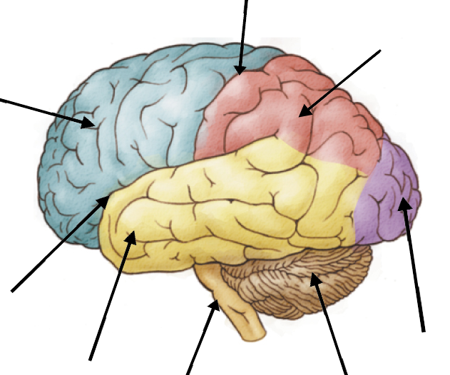
The cerebral cortex is composed of four distinct zones, or lobes; because the nervous system is bilaterally symmetrical, we have four paired lobes: frontal, parietal, occipital, and temporal.
Rotate the figures below by either moving the image with your mouse/touchpad or using the arrow buttons. The whole brain image on the left allows you to examine the outer surface of the brain, and the half brain image on the right shows a side view of the brain’s central structures. Select the each various regions of the cerebral cortex to learn more about each cortical lobe.
Identify the various brain structures by matching the colored circle on the drawing with the correct term.
The interior of the brain is not homogenous but is instead made up of dark and light regions, commonly referred to as gray matter and white matter, respectively. Gray matter is largely composed of cell bodies (somas) and capillaries, and it is typically where neurons collect and modify information before sending it elsewhere. White matter is mostly axons covered by myelin, the fatty substance that gives these regions a white appearance. White matter forms longer-distance connections within the nervous system.
Select different cuts to view brain slices in three different orientations. Roll over different areas of each brain section to distinguish gray matter from white matter.
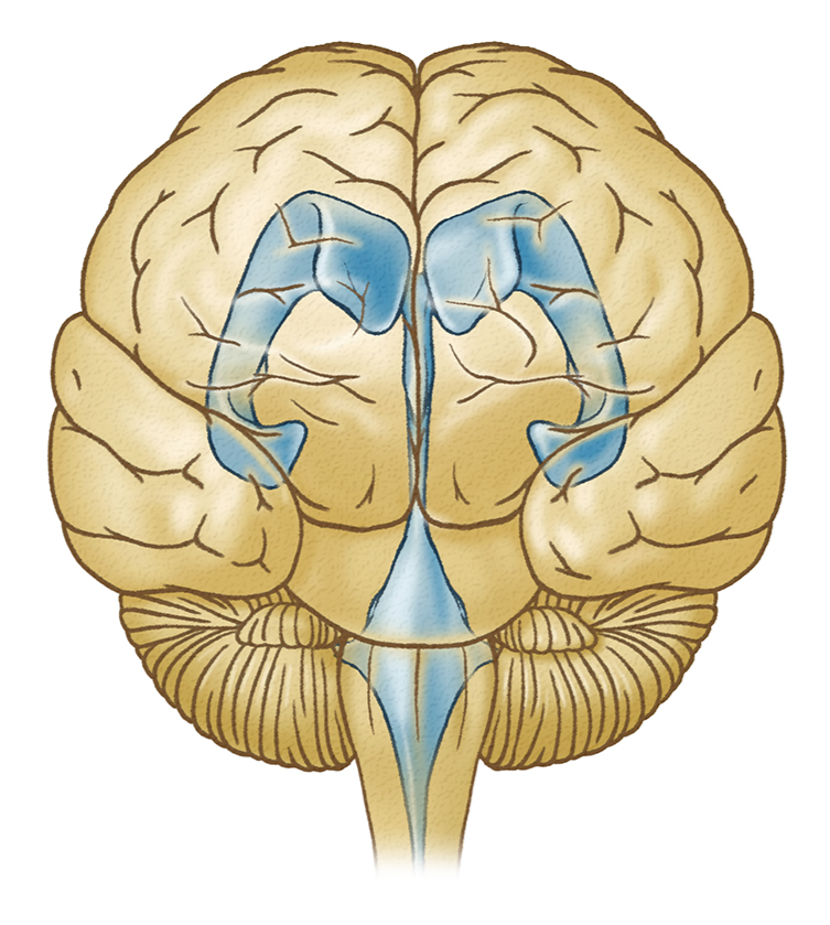
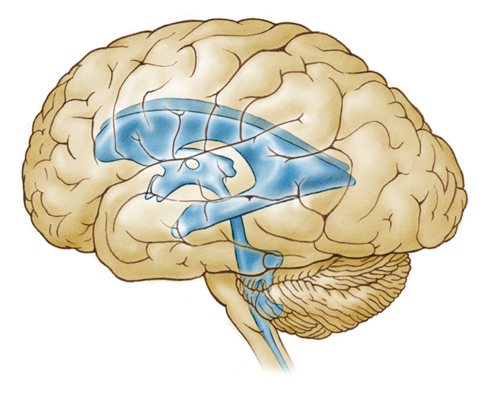
The brain’s interior also contains four interconnected fluid-filled spaces, called cerebral ventricles, or ventricles. Cerebral spinal fluid (CSF) circulates through the four ventricles: two lateral ventricles, the third ventricle, and the fourth ventricle (CSF flows from the two lateral ventricles, called the lateral aperture and lateral ventricle, into the third and fourth ventricles, and then into cerebral aqueduct which runs the length of the spinal cord). The flow of CSF through the ventricles allows nutrients and other important substances to be delivered to brain cells and for waste products to be carried away.
Explore the interconnected system of cerebral ventricles in three dimensions. Rotate the figures below by either moving the image with your mouse/touchpad or using the arrow buttons.
Question 1.
P5y/3noc1PGRanXfXE4SAR7H4WXLOJiAHPEwvU6/raDiGqCn/bne2zIvrMD/xzMmuMM2c2XS9slfONs0sUNQFqjmudI7tlG7HT75Aviy/VA85/tA6B3PS+ZAnI0mpDoeciM9n04+puR0KKsT1JWkBRIxGK/b9EqOYour answer has been provisionally accepted. You'll get full credit for now, but your instructor may update your grade later after evaluating it.
Item 2 of 2: Match the labels to the ventricles.
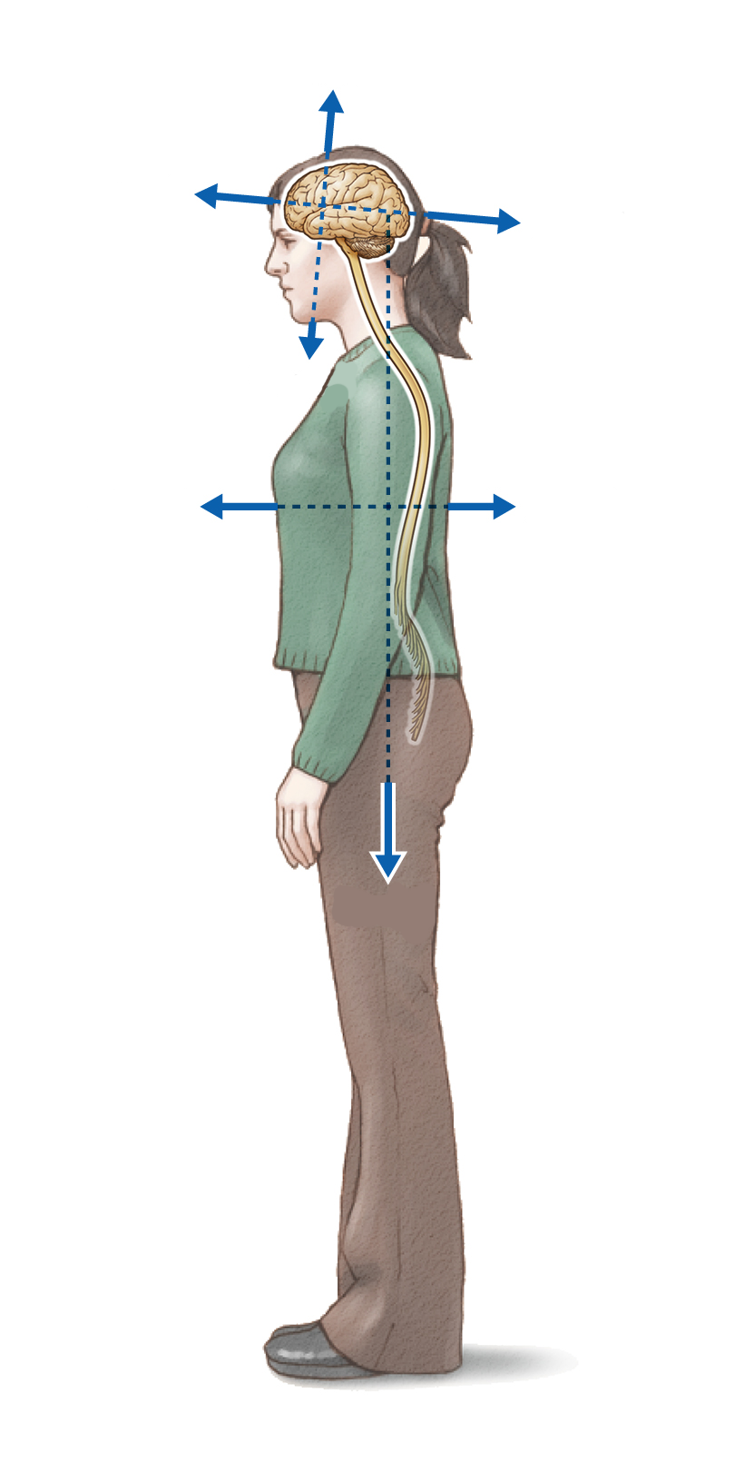
A variety of directional terms are used to describe the position of nervous system structures, either in absolute terms (for example, the ventral striatum) or with respect to the position of other anatomical structures (for example, area A is ventral to area B). These terms provide a common language for scientists to describe the location or position of nervous system structures.
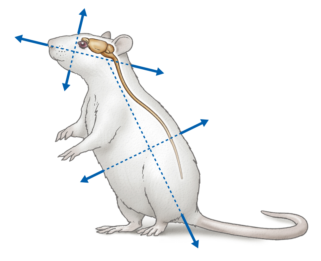
Select each arrow to learn how these directional terms are applied to a quadruped (in this case, a rat).
dorsal ventral rostral caudal
rostral caudal medial lateral lateral
In rats and other quadrupeds, the brain and spinal cord are parallel. But when rats stand on their hind legs, the spinal cord is no longer parallel to the brain; instead, it is rotated nearly 90 degrees. However, despite the change in the spinal cord’s orientation from horizontal to near vertical, the directional terms remain consistent.
To apply the directional terms to a rat standing on its hind legs, match the labels to the arrows.
Note: you must choose all answers before proceeding.
Question
Question
Congratulations! You successfully completed the Finding Your Way Around the Brain activity. In this activity, you reviewed the brain’s major features, both internally and on the surface. You also reviewed directional terms commonly used to describe the positions of nervous system structures.
Your instructor may now have you take a short quiz about this activity. Good luck!
