Chapter 1. Visual Pathways: The What and the How
Visual Pathways: The What and the How
By:

We often think of using vision as a tool to recognize objects, words on a page, or familiar faces, but we use our vision for more than just simple recognition. In this activity you will learn about the brain's visual processing system by studying the two major visual streams outside the primary visual cortex: the dorsal stream and the ventral stream.
After completing this activity, you should be able to:
- Explain the roles that the dorsal and ventral streams play in the processing of visual information.
- Describe how injury to the visual pathway can lead to visual disorders.
This activity relates to the following principles of nervous system function:
- Principle 1: The Nervous System Produces Movement in a Perceptual World the Brain Constructs
- Principle 4: The CNS Functions on Multiple Levels
- Principle 6: Brain Systems Are Organized Hierarchically and in Parallel
- Principle 8: The Brain Divides Sensory Input for Object Recognition and Movement
- Principle 9: Brain Functions Are Localized and Distributed
Visual information travels from the eyes to the brain. After passing through the thalamus, the information enters the primary visual cortex (V1, or striate cortex), which is located in the posterior occipital lobe. From there, information can go into one of two streams: it can go through the dorsal stream, also called the “how” pathway (toward the parietal lobe), or through the ventral stream, called the “what” pathway (toward the temporal lobe).
This activity demonstrates how different information is processed in each of these streams. But keep in mind that these two streams can pass information to one another, so they are not completely independent.

The dorsal stream is the pathway from the occipital lobe to the parietal lobe, and the ventral stream is the pathway from the occipital lobe to the temporal lobe.
Based on the results from patients with visual deficits, David Milner and Mel Goodale proposed that these two visual streams are responsible for two distinct functions: identifying a stimulus (the "what" function, which is a product of the ventral stream) and controlling movement toward or away from the stimulus (the "how" function, which is a product of the dorsal stream). Patients with localized damage to one stream or the other have very specific visual deficits, as you will soon see.
Before we meet our patients, let’s review the functions of the two visual pathways. Information arriving in the primary visual cortex can follow two streams: What are these two streams, and what does each stream do?
Question 1.
6P3D/G7/3G9LkNzr1iaYRZr9PuC49SaiYyupI0MOGqcTVOgpAyh8FtROUJcQe6SXNE1pc7siF992OwCCg84ckc7V8g9LIAGFeZ/0rzEQFPlT36FFRYWT3nCPsIC4mdVdfxi9iFQW9kLa/nwSNYQqyY1lJrKAAD0gR0BqG/Y+7Gh4mMQNmNcaN7U/zOUqRgdqnqPfT5cde/oBnscMwj31lFXR1GeUH1hvGdJvygfJcFkt67+vBro9E5qP3MvntJJDxERbKbaXJdalGF5mInN8HZWaey1aNGe3cxSzEy1+Ca8Fi5Yx6WoihSvOhxeINg6WgJhl2OmMM9K1Bn2gTBgknPwngcYHgfmfYour answer has been provisionally accepted. You'll get full credit for now, but your instructor may update your grade later after evaluating it.
Let’s meet two patients who have sustained selective, localized brain injury to visual areas of the brain. Click on each patient’s name to discover more about her injuries.
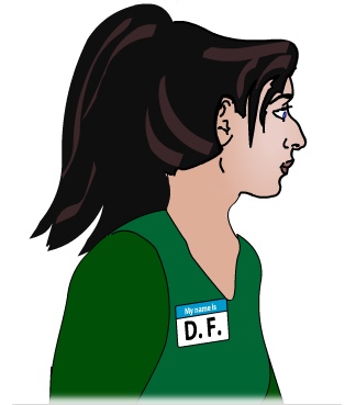
Patient D. F.
Patient D. F. was 35 years old when she sustained accidental carbon monoxide (CO) poisoning. It is unclear how long she was exposed to the CO, but the exposure resulted in extensive damage to her lateral occipital region in both hemispheres.
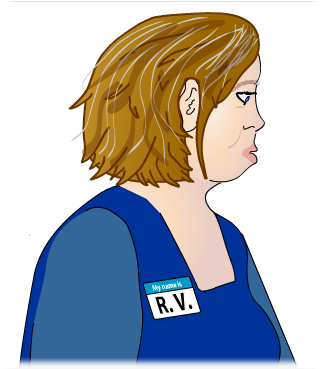
Patient R. V.
Patient R. V. was 55 years old and had sustained a series of strokes. The result was extensive damage between her occipital cortex and parietal cortex in both hemispheres.
Patient D. F.: Goodale, M. A., Milner, D. A., Jakobson, L. S., & Carey, J. D. P. (1991). A kinematic analysis of reaching and grasping movements in a patient recovering from optic ataxia. Nature, 349, 154-156
Patient R. V.: Milner, A. D., & Goodale, M. A. (2006). The visual brain in action (2nd Ed.). Oxford: Oxford University Press.
Question
q6XWQJflWtVkaFwYK6NEneWW7Y2E1zzqMCTXSg==What happens when the areas of the brain that are responsible for visual processing are injured? We can answer this question by studying patients who have damage to specific cortical areas.
The following tasks are commonly used to evaluate visual deficits. For each task you will evaluate how patients D. F. and R. V. perform.
Note: you must evaluate all tasks before proceeding.
Task 1: Object recognition
This simple recognition task asks the patient to identify a number of common objects using only her vision (that is, she is not allowed to pick up the object or gather any other sensory information from the object). The patient must therefore be able to visually process details of each individual object and then identify it.
Was the patient able to recognize the objects?
| D.F. | R.V. |
|---|---|
Questionfy5YUOMiClGw7NjrO6G3TcduRnQnbWT0vgs7gbsMG6K1kupFUd/8ocBkRisl8q2nHdnrUsbFmkVbIoHF/4EGAIl367GluOssDccoAYixf31kRheT+cXp5JYCQ4qCvk0RoNs4oI7tOjgpAtJvklewublzrAlyHobrE9yTjLBAa0Lx7khxaX0HEiUw8ZG7Bbqu9LR3jamo4H+HwDT9Tbn1M6kPlyBPpHuqow9Vmnb1ULRSPRUW/bPw5w==true
true
true
{qqMC1 === 0} setModel("one_1", "yes")
{qqMC1 === 1} setModel("one_1", "no")
|
Question2J0sMncAdaKywr7c28C4z1FT8uejEaKSeLmJwudbgLvv/uogIKc2IrjFZmC+lvnMQX/yl6UxS8RJBkczml6SE4352kwEMDbAEvBGkbPKT+yCTL8gesQtiCC9J9ADqtSKuMlKUIUwjGeYW2qHR4C7KWktevJUtxuTIBwIJCAvAkgMgGnpPE7ohzBJw8g5kwnjVJUwefBjQqTPHwxPr9/nhi7ld/B08H9mjBPvCVztA8MtahX24xvghQ==true
true
true
{qqMC1 === 0} setModel("one_2", "yes")
{qqMC1 === 1} setModel("one_2", "no")
|
Task 2: Line copying (drawing)
In this task, patients must copy simple line drawings on paper. In this case, the patient is given drawings to copy, but this task can also be done with models of the objects. This task requires the patient to be able to visually process details of the line drawings and copy them on to a separate sheet.
Was the patient able to copy the line drawings?
| D.F. | R.V. |
|---|---|
Question6V3dlzz8H+4Bg/CSFxkIRkykZ06dpVLklJZJLvM9CB8vwpiATMJ7pySU/WszuYgw7Q60/ttdjB/cyNCeFszqggEfVnpHuttistP1jNx7uLpaMwZsXfzXW3Flh38aD75hnoAO5oYRE4IfIEDPo/x1H9YUajHJZUQhUk2CJSxUvJqZgMLpQM4lLNm9YdaM64gTftsdYrvgPzwcVqCnvrwO58nGUhTcsJqE4FaPRP0LIFjDDGVhwIv7Cg==true
true
true
{qqMC1 === 0} setModel("two_1", "yes")
{qqMC1 === 1} setModel("two_1", "no")
|
QuestionwPJOe8HlnPCrMC/RYHXQbC6gXbFM5RHTMG79S4ds38P8hIH+J1YdTmio1cziyEve59hYWGdIXzXkGMnCI7h9HB+GoKCz/cDoP4uEgAAh1MvS4H0LvdWeqF/o/ATlTgin8JoFDmX/zGV4vv2Cu0yRb+gWFE3NJLLh+ptHp+2xtGDIDJHH2wIi47ivgtTmxjJg0r1m+DK+4O/+47N6/9m6AxbjVm9fGA9tRar6p/RGyQsHO0EZvo4OIg==true
true
true
{qqMC1 === 0} setModel("two_2", "yes")
{qqMC1 === 1} setModel("two_2", "no")
|
Task 3: The Matching Task
In the first component of this task, known as the matching task, the neuropsychologist has asked the patient to match her hand to the orientation of a slot. Notice that this requires the patient to be able to use her visual perceptual system to determine the orientation of the slot, but it does not require any motion toward the slot. The neuropsychologist can change the orientation of the slot for subsequent trials.
Was the patient able to match their hand to the orientation of the slot?
| D.F. | R.V. |
|---|---|
QuestionrekK1VyiqCnZTm2A38pXCz8dPDFDEBHgnKZwDLiSuTTyJBn9MGJYOQtPobJ4E+CbjhX5mdRjazLb9PSpyqcQO8UqKYjpAJHenHkQZf8JkSzPqufBRgNk4J4DBfhPPFnmTuU2bOUQoFDvJRCt3zRyngVeE1AVpD6ovd84RkjAoRGHRMsgG12HrfS34O/DFGEt6FOWqPBlKuIY91uBf5A2Qo7VlVU5KMHmOJtZbupKvMLYMUf+fPBstdV/j3cap1iFs2Iv3B+9gBoV493pkjx8YQ==true
true
true
{qqMC1 === 0} setModel("three_1", "yes")
{qqMC1 === 1} setModel("three_1", "no")
|
QuestionAQvCxJakg0Xc1mM3sel5zTvLHPcAv3pFvzikDF5oR7aNdcYGxw/1nSAUle9pd3T2vj5UDg/fG556i3dK0TZsoX6XWq/FZSuHGbt5JqpZJAtlzszM6XTTS1P5UqdyOl6MNUpCABDast+GPG9F/nttJdKaa9cWfXCgDr4kpaeFHIWlqivGfwbr2ZftgDf6Qx55+kAI4sVWEGv7L9EguCvwN5ZLFNzQ33YUmek11OuQrZ2+wU0IZhuTkmLYOV8CGzkC94QAwDdBxSBJtYzgK4crTw==true
true
true
{qqMC1 === 0} setModel("three_2", "yes")
{qqMC1 === 1} setModel("three_2", "no")
|
Task 4: The Posting Task
In the second component of this task, known as the posting task, the neuropsychologist has asked the patient to post a card and post it through the slot, as if it were a mailbox. This requires the patient to be able to use her visuomotor system to guide her actions as she posts the card.
Was the patient able to successfully post the card in the slot?
| D.F. | R.V. |
|---|---|
QuestionRgZkswnhPb9kA/T6ZwZubLo10gvC7+HcMiwhdPdoyunNvq8YMS2wBdX8F7Q5fAObS7j5ROF4XFupgeMatlJww55JQCdDxXe+ahYoDpYWo75cljONqeconTHMVuGE/JL3dEddn9Xyi19Qmr7wuuNumHQQ/jWQnH5T4W/EG+xFWoMxQVbHptWX6fKzqGsgb1A1+9BTOkHB6cprvFtEpGynOzJqj1fXAuGzzFXWHky1x9o4OIctoT2IYtbCp1MDzwcAVT+SaUZUlN4=true
true
true
{qqMC1 === 0} setModel("fourth_1", "yes")
{qqMC1 === 1} setModel("fourth_1", "no")
|
QuestioncOTRlp3lf19xXxSexUbIiuAMd7ujWWgv6rkttugsOE3Jyh3D9tOdqt0v+uY4/hIb6lBzDVi2gkujq73UL5W7OkyyGDEXYJkHPqDTN7CA5zwuzfyFr7UbShKUynXNTZpiGPvte0XVbY2h7hAI5vgTOJjB5otYlQOznLnJ73JnXHCldB0vUu3PXSznD+sVPRH+szu739MFv2SCPEF1GTS7GcmvCw6U9nuxK7r0H5XWVDRGm1wB9MSngU6sUpO+gs3S1wgRmIgIqSY=true
true
true
{qqMC1 === 0} setModel("fourth_2", "yes")
{qqMC1 === 1} setModel("fourth_2", "no")
|
Task 5: Grasping task
A grasping task can be used to evaluate whether a patient is able to use vision to correctly adjust her movements. Notice that the hand shape varies considerably depending on the object that is being grasped: to pick up the pen, the fingers must be configured in a pincer grasp, but this type of grasp would not work well for picking up the coffee mug.
Was the patient able to grasp the objects correctly?
| D.F. | R.V. |
|---|---|
QuestionPSK0WoqUK6Ig36s7PyTAU4RcqWHVEJ5X/syOz4ipu+vhCG+737HW8jL0KgJitqkrwE3qVzYeSrZKG6Tn/PFzDm3IN9EJot2zHMuifk+nKnl8YnmX5TJsPVuroFtSA0092igwNoHWmtIbGfnHCBfPokD7TqGwseDTK25KFTSdxzTebWVyv8/diFWPLLLTg/sfuBbdVcbpt/lKY1yD9vURrUud+kEUr5X8O8rdutDESsz3zDCITlLdn7aphao=true
true
true
{qqMC1 === 0} setModel("five_1", "yes")
{qqMC1 === 1} setModel("five_1", "no")
|
QuestionnCCEInHH4il2AmDKpEExJGn7z0lEaEMWMYN85g3w8jP+qHIA4z36j70xHz4J75hm6HykW2z0qVK4vA+j9EAInO38v51bWDeOy/TvGRcO4a4gqpmGCUNOOqqEWc6x8nsBifDF6iO1xL9iREVIQXgeEpW7d7ZFeZIo+kVWkhMdDlbQf0j3D9FRW13LkiBFVRJ6zeO6ZL0nYlHoM31BEkeyUqNQjcUzYAiGowDTXH0yS2bv8LiNvJw1HaAmUCU=true
true
true
{qqMC1 === 0} setModel("five_2", "yes")
{qqMC1 === 1} setModel("five_2", "no")
|
The results of Patient D. F. and Patient R. V. (shown in the table below) differed considerably on each of these visual tasks. Note that for both of these patients, visual deficits were caused by selective injury to a portion of the visual areas of the brain; their eyes remained healthy and functional.
| D.F. | R.V. | |
|---|---|---|
| Task 1: Was the patient able to recognize the objects? | no | yes |
| Task 2: Was the patient able to copy the line drawings? | no | yes |
| Task 3: Was the patient able to match their hand to the orientation of the slot? | no | yes |
| Task 4: Was the patient able to successfully post the card in the slot? | yes | no |
| Task 5: Was the patient able to grasp the objects correctly? | yes | no |
Question 2.
bZ1iaY4do8umNIGAzjmcTBW/p4bczhM/UiOa6QKFXQLDIBd78FbkCw1LK+/N8ZWXHLzhRAAyKe0VsLjGyzpjAdAXQ50yIiAG0vNeX/gPeVKfGEocvNugveXzv5qBZNufHN027A7+0B+REXKrSeFZwFvYH5oJ6Hz8X+BBrT5hWmmyw5VOooAoWt9swls1fcMXu/ucDseccusUL2d54gId+1vJX2/oCVzd4mQp7xS2tqHSTAD3B1a5OfqmAJWkIQYcLMmDLn+0q6jIGFkBpvoSZWjKm2y1zb4Vu5RecmaGsJe3tuGIq8wXv5sec50o+BDuvVYMJB5BFGLKLmqVQAl1BgLCAgKmvauIWD2NwzaoaoYZMpzPYour answer has been provisionally accepted. You'll get full credit for now, but your instructor may update your grade later after evaluating it.
The specific visual deficits experienced by D. F. and R. V. demonstrate the unique functions of the brain’s two visual streams.

Patient D. F. has selective damage to her ventral stream. She has a condition known as visual form agnosia. This patient cannot use vision to properly identify and recognize objects but can use vision to properly guide actions. This caused her to have some strange visual deficits: while she could not perceptually match her hand to the correct orientation in the posting task, she could use her vision to correctly guide her movements in the posting task and in grasping objects.

Patient R. V. has selective damage to her dorsal stream. She has a condition known as optic ataxia. This patient can use vision to properly identify and recognize objects but cannot use vision to properly guide actions. In the grasping task, she was not able to guide her fingers into the necessary position to pick up the various objects.
Congratulations! You have successfully completed the activity. In this activity, you learned about the roles that the dorsal and ventral streams play in the processing of visual information and how injury to these visual areas can lead to specific visual deficits.
Your instructor may now have you take a short quiz about this activity. Good luck!