Chapter 1. LAB 7 Laboratory Skills 3: Dissection
Introduction
Learning Goals:
- Learn dissection technique as a skill and method for investigations into animal structure and function
- Understand the complexity of bilaterally symmetrical invertebrate and vertebrate animals by comparing their structures and the ways in which they function
Lab Outline
Activity 1: Demonstration of the Dissection Strategy
Activity 2: Earthworm Dissection
Activity 3: Dissection Specialization and Investigation
Scientific Inquiry
Many animal species alive today are unknown to us. What are they, and why should we care about them? Why not focus only on humans? Animals play many profound roles in the world’s ecosystems as members of food webs and agents that control pest species, as well as posing benefits or threats to human health (e.g., parasites). Because our well-being is so intricately connected to other living things in this world, it is in our best interest to understand the ways in which they contribute to the web of life. This understanding requires scientific investigations of organisms at all stages of their life, in their natural habitats. Such investigations open up a fascinating world of new insight into the living things around us.
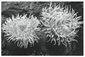
Recently, with the aid of molecular techniques, we are also discovering how intricately our ancestry is linked to all other members of the animal kingdom. In the July 6, 2007 issue of Science, Putnam et al. reported the surprising similarity to vertebrates of the genome of one of Earth’s oldest animals, the sea anemone (Figure 1). The authors analyzed DNA from the starlet sea anemone and compared it with that of other animals. They found that two-thirds of the gene families in humans and sea anemones are derived from an ancient common ancestor referred to as the “ancestral eumetazoan.” Previous research relied heavily on animals such as fruit flies and worms to study our relationships to other living things, but many of these organisms have lost the genes from the ancestral eumetazoan, whereas the sea anemone has retained these ancient genes, just as humans have.
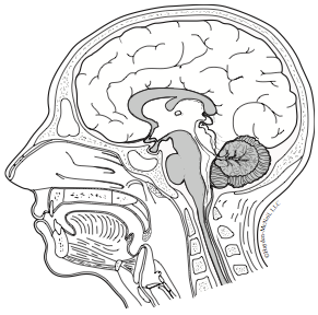
Modern biologists can collect molecular data that informs us about our ancestry so we can make important inferences about evolutionary relationships among organisms, but biologists are also interested in the basic structure and function of the anatomy of individual animals and use this knowledge to make new technological advances. For example, applied knowledge about the anatomy and sensory functions of the brain, eyes, and the tongue (Figure 2) has led to the invention of brain computer interface (BCI) technology that allows the blind to see with their tongue (Ptito et al. 2005; CBS News 2007; Danilov and Tyler 2005). Yes, you read that correctly—with this technology, the blind can use their tongues to see. The subjects wear a camera that receives visual signals, functionally replacing the eyes. The visual signal from the camera sends electrical impulses to a series of electrodes situated on a pad that is held on the surface of the tongue in the mouth, a TDU (tongue display unit). The tongue is electrically stimulated by the signal sent to the TDU electrodes and naturally sends a signal to the tactile center of the brain. Amazingly, over a short period of time “cross-modal plasticity” occurs: the brain rewires itself so that tactile signal is rerouted to the visual cortex allowing the subject to sense what the camera is viewing. How is this possible? The tongue, which is normally wired to the tactile sensory areas, is serving as a conduit or passageway for getting the visual information to the visual cortex of the brain (Ptito et al. 2005; Danilov and Tyler 2005). The tongue switches from a tactile sensory mode to a visual sensory mode in communicating with the brain, hence the term “cross-modal plasticity.”
As you explore representative members of the animal kingdom through dissection, think about the similarities and differences between their anatomical structures. Develop an appreciation for the scientific knowledge acquired through basic dissection and the further advances that have developed state of the art noninvasive technology like the TDU device. Make connections between structural form and function as you do this and you will discover how they relate to an organism’s natural and evolutionary history, as well as how systems can be manipulated for human advancement.
Background
Basic survival of multicellular organisms depends on the delivery of adequate sources of nutrition, gases, and fluids to all cells in the organism, as well as elimination of waste metabolites, gases, and fluids from all cells in the organism. Plants achieve these functions using diffusion and vascular tissue in their four primary plant organs: roots, stems, leaves, and flowers. However, animals are more complex; they don’t make their own food like plants do, so they must obtain nutrients from their environment. Also unlike plants, animals generally have to move, if only to capture food, so they need body structures that facilitate locomotion. While single-celled organisms perform all the functions needed for life within their cells, metazoans divide the workload of locomotion, nutrition, metabolism, and elimination into different cells, tissues, and organs. Only the exterior of the animal’s body contacts its physical environment, while the cells, tissues, and organs responsible for maintaining metabolic homeostasis are generally enclosed within the body wall. To understand these structure–function connections, we have to dissect the organism.
Dissection is the practice of closely examining a three-dimensional object and all of its layers in order to discover its parts, understand their functions, and in some cases repair parts that are no longer functioning the way they should. When one considers the diversity of animals, dissection would appear to be a rather complicated process. Fortunately, the stunning diversity of animal forms are actually built according to relatively few body plans, so the understanding of how to dissect one kind of organism can be applied to other kinds of animals. In dissection of animals, the body wall is opened and the connective tissues holding the organs in place are pulled aside so one can look more closely at the structures and connections between the organs. When the organs are opened, one may look more closely at the tissues and cells that make up the organs. Details of tissue and cellular structures reveal possible locations for the biochemical machinery that drives cellular function.
In biology research labs, dissection is often necessary to access an organ, a tissue, or a cell of interest from an intact specimen. The first challenge in correctly dissecting an organism is the ability to perceive it in three dimensions. This is not an entirely novel task, even if you have never “officially” dissected something before. We perceive three dimensions in our heads in order to perform many day-to-day tasks. Have you ever tried to assemble a new piece of furniture or a new toy with a picture diagram? The illustration obviously helps, but we cannot truly understand or perceive 3-D objects from a flat image. Have you ever tried to cut a piece of pie without slicing into the pie tin that holds it? This requires depth perception, as do commonplace activities like driving a car and playing sports. Use your skills of depth perception and translation to interpret a two-dimensional image of a three-dimensional object so you can dissect and analyze parts of different animals. In doing so, you will gain appreciation for the skills a surgeon must have, as well as develop your own ability to mentally build 3-D images from 2-D drawings.
Resources
1.1 Lab Preparation
Lab Preparation
Watch the vodcast and read this lab. Write all notes in your lab notebook.
Activity 1: Demonstration of the Dissection Strategy
Learning Objectives
After successful completion of this activity, you should be able to:
LO15 Demonstrate proper use of one or more of the following tools: Vernier caliper, micropipettors, digital balance, graduated cylinder, spectrophotometer, pH meter, and compound and dissecting microscopes
LO74 Dissect and reveal meaningful features of representative organisms or parts of organisms using appropriate dissection instruments
LO75 Think and manipulate in three dimensions using fine motor skills
Basic Dissection Safety: For your safety and the safety of your classmates, thoroughly review the general dissection strategy guidelines that follow before you begin dissecting. Practice safe hygiene. Wear gloves and thoroughly wash all utensils and your hands when you are finished. Dispose of waste in the proper receptacles. DO NOT eat or drink anything while doing dissections. Do not bring food into laboratories.
Activity 1 Procedure
- During the dissection demonstration, complete the application column of the dissection strategy (see below) in your lab notebook. For example, if you were dissecting a clam you might state in the application column next to “Examine external features” that the clam has two light-colored, hard shells with circular striations. The shells are held shut by muscles. There is a bulge in the clam shells near where the two shells are attached called the umbo.
- Make sketches, ask questions, and take notes on the dissection technique and the organism as the demonstration proceeds.
- Share your findings and helpful hints with your teammates.
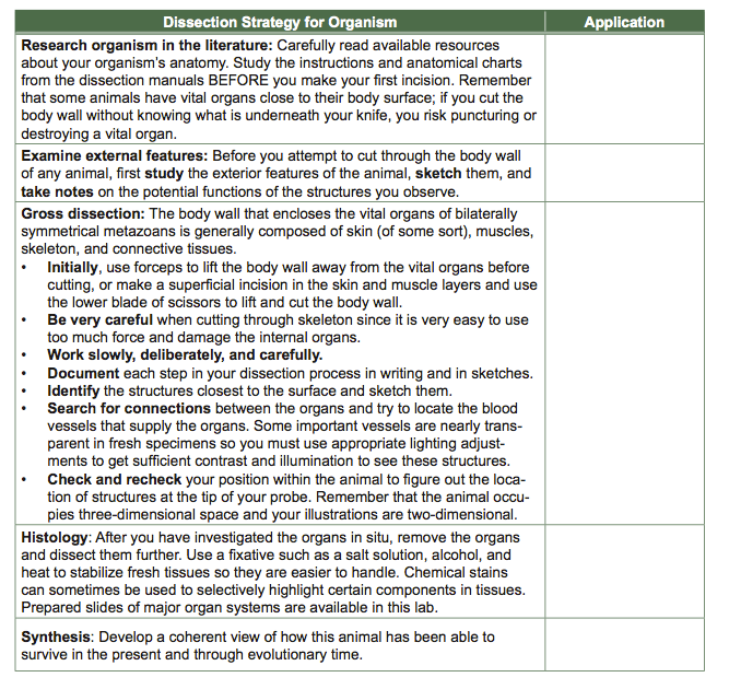
Dissection Strategy Helpful Tips:
- Use blunt-edged tools such as the dissecting probe before using pointed tools as they are more forgiving and are less likely to cut the tissues than a scalpel or needle. Blunt-edged tools should always be used for separating tissues, organs, glands, etc., from each other.
- As a general rule, use scissors before using a scalpel: you will have more control over your cut. When a scalpel is absolutely necessary, for instance to make a primary incision, complete the cut with scissors, using care to only cut the appropriate tissues.
- Prevent desiccation by using water or saline: most organs are easier to see when immersed in water— once you have exposed the body cavity, pour water slowly into your dissecting tray until the internal organs are submerged.
- Expand collapsed organs: immerse in a solution to make the organ float, or inject organs with water, air, or plastomer.
Activity 2: Earthworm Dissection
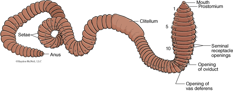
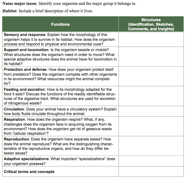
Use the species-specific dissection instructions in the dissection appendix (Appendix D). As you work, focus attention on the listed basic information about the organism and its functions. Write your answers in a dissection summary table in your lab notebook. You are required to sketch the anatomy and write about it, but you may also make a record of your dissection using a digital camera (optional). For this portion of the lab, work in pairs to dissect and closely study the gross anatomy of an earthworm.
Learning Objectives
After successful completion of this activity, you should be able to:
LO76 Open the body wall of a soft-bodied animal without damaging underlying organs
LO77 Separate the organs without damage, by releasing them from the connective tissue layers that bind them together
L078 Identify the major organ systems and the individual organs in the earthworm including the circulatory, excretory, digestive, and nervous systems of the earthworm
LO79 Recognize the layers of the body wall in cross section including the circular and longitudinal muscles
LO80 Describe structures and their functions in animals
Dissection toolkit
Disposable gloves
Your dissection organism
Water (for organism immersion)
Dissecting scope
Activity 2 Procedure
Work in pairs. Get a dissecting pan, pins, and a dissecting scope with lamp for your group.
- Retrieve an anesthetized worm from the dish (Figure 3 and Appendix D for external anatomy). Apply the dissection strategy for your earthworm dissection, with help from the instructions in Appendix D.
- Find and carefully trace the relationships among the structures in your organism. Create a table in your lab notebook of the structure and function of the major organ systems (see the template below).
- Take notes and sketch these structures to clarify your understanding of their connection and relationships within the body cavity. Explain how you identified these structures and how these structures function in the context of the animal’s lifestyle.
Activity 3: Dissection Specialization and Investigation
Learning Objectives
After successful completion of this activity, you should be able to:
LO81 Compare important functional features of organs with similar functions in other organisms and how these structures perform their functions given the organism’s lifestyle
LO82 Successfully teach other individuals what you have learned about your assigned organ system in different animals
Activity 3 Procedure
- Your lab instructor will assign you and your lab partner a particular Exploration Module (below) in which you will explore organ/organ systems in vertebrate(s) and invertebrate(s).
- Refer to the species-specific instructions in Appendix D to find out how to dissect your animal and organ system. Groups will work together on the same rodent, so complete your dissection carefully, being mindful to leave the other organs and organ systems intact for dissection by other students.
- Discuss each organ as a group of four students and take notes in your lab notebook. It may save you time to construct a data table with structure, function, and the organisms dissected in this lab (earthworm, rodent, etc.) as column headings.
Exploration Modules: System to Organs to Tissues to Cells
- GI (gastro-intestinal) tract of the earthworm, rodent, and fish. What organs make up the GI system in these animals? How does each animal gather food, digest its food, and absorb the nutrients? Open the different sections of the GI tract and compare the tissues lining their cavities. Use prepared slides to see their structures in cross section.
- Excretory (kidney) systems of the earthworm, rodent, and sea star. What organs make up the excretory systems of the rodent and earthworm? Where are the nephrons in the earthworm and rodent? What happens to the excretory products after they leave the nephrons in both animals? Use prepared slides of the gross kidney anatomy and the earthworm nephron.
- Circulatory system of the earthworm, rodent, and reptile. What organs make up the circulatory systems of the rodent and earthworm? Dissect a portion of the aorta and the posterior vena cava. Compare the structure of the walls of the artery and vein. Use pre-prepared slides to see these structures in cross section.
- Respiratory systems in the earthworm, rodent, and clam. How is oxygenated blood transported to the entire body in each animal? What major specializations does each have so it can survive in its habitat?
- Reproductive systems in the earthworm, rodent, and frog. What structures make up the reproductive systems in these animals? How does fertilization occur in each animal? Where do the fertilized eggs develop?
- Musculo-skeletal systems in the earthworm, rodent, and crayfish. Why do mammals need bones in order to move? How do earthworms and clams manage to move without hard skeletons?
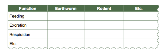
Anatomical Comparisons Presentation
- Prepare a data table in your laboratory notebook similar to the one below. Draw a table with the organisms that the class dissected in Activity 2 and 3 as column headings, and the principle functions (e.g., feeding, excretion, respiration, etc.) as row headings. The data table is designed to help you organize the structures you have just learned to make their structures and functions easier to compare.
- Fill in the appropriate structure(s) for each function under the heading for each organism. To ensure that you remember defining features, include sketches of the organs from your lab notebook, their position in the animal, and their role in life functions. Label your sketches.
- The class groups will teach each other about their organism and its anatomy, and provide insights into the unique ways each organ functions. This is a good time to ask questions and deepen your understanding of the animal, its anatomy, and its lifestyle.
- Fill in your comparison table. In what ways is this organism similar to the organism you dissected? What aspects of the organism are unique?