Chapter 1. Impact 14.1
Impact…ON MEDICINE: I14.1 Magnetic resonance imaging
One of the most striking applications of nuclear magnetic resonance is in medicine. Magnetic resonance imaging (MRI) is a portrayal of the concentrations of protons in a solid object. The technique relies on the application of specific pulse sequences to an object in an inhomogeneous magnetic field.
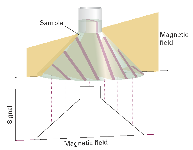
If an object containing hydrogen nuclei (a tube of water or a human body) is placed in an NMR spectrometer and exposed to a homogeneous magnetic field, then a single resonance signal will be detected. Now consider a flask of water in a magnetic field that varies linearly in the z-direction according to \(\mathcal B\)0 + \(\mathcal G\)zz, where \(\mathcal G\)z is the field gradient along the z-direction (Fig. I14.1)). Then the water protons will be resonant at the frequencies
\(\nu_{\mathtt{L}}(z)= \frac{ \gamma_{\mathtt{N}} }{2 \pi } ( \mathcal {B}_{0} + \mathcal {G}_{z}z)\)
(Similar equations may be written for gradients along the x- and y-directions; the L denotes a Larmor frequency.) Application of a 90° radiofrequency pulse with \(\nu= \nu_{\mathtt{L}}(z)\) will result in a signal with an intensity that is proportional to the numbers of protons at the position z. This is an example of slice selection, the application of a selective 90° pulse that excites nuclei in a specific region, or slice, of the sample. It follows that the intensity of the NMR signal will be a projection of the numbers of protons on a line parallel to the field gradient. The image of a three-dimensional object such as a flask of water can be obtained if the slice selection technique is applied at different orientations (see Fig. I14.1). In projection reconstruction, the projections can be analysed on a computer to reconstruct the three-dimensional distribution of protons in the object.

Fig. I14.1 In a magnetic field that varies linearly over a sample, all the protons within a given slice (that is, at a given field value) come into resonance and give a signal of the corresponding intensity. The resulting intensity pattern is a map of the numbers in all the slices, and portrays the shape of the sample. Changing the orientation of the field shows the shape along the corresponding direction, and computer manipulation can be used to build up the three-dimensional shape of the sample.
In practice, the NMR signal is not obtained by direct analysis of the FID curve after application of a single 90° pulse. Instead, spin echoes are often detected with several variations of the 90°–τ–180° pulse sequence (Topic 14C). In phase encoding, field gradients are applied during the evolution period and the detection period of a spin-echo pulse sequence. The first step consists of a 90º pulse that results in slice selection along the z-direction. The second step consists of application of a phase gradient, a field gradient along the y-direction, during the evolution period. At each position along the gradient, a spin packet will precess at a different Larmor frequency due to chemical shift effects and the field inhomogeneity, so each packet will dephase to a different extent by the end of the evolution period. We can control the extent of dephasing by changing the duration of the evolution period, so Fourier transformation on τ gives information about the location of a proton along the y-direction.* For each value of τ, the next steps are application of the 180° pulse and then of a read gradient, a field gradient along the x-direction, during detection of the echo. Protons at different positions along x experience different fields and will resonate at different frequencies. Therefore Fourier transformation of the FID gives different signals for protons at different positions along x.
A common problem with the techniques as described so far is image contrast, which must be optimized in order to show spatial variations in water content in the sample. One strategy for solving this problem takes advantage of the fact that the relaxation times of water protons are shorter for water in biological tissues than for the pure liquid. Furthermore, relaxation times from water protons are also different in healthy and diseased tissues. A T1-weighted image is obtained by repeating the spin echo sequence before spin–lattice relaxation can return the spins in the sample to equilibrium. Under these conditions, differences in signal intensities are directly related to differences in T1. A T2-weighted image is obtained by using an evolution period τ that is relatively long. Each point on the image is an echo signal that behaves in the manner shown in Fig. 14C.14, so signal intensities are strongly dependent on variations in T2. However, allowing so much of the decay to occur leads to weak signals even for those protons with long spin–spin relaxation times. Another strategy involves the use of contrast agents, paramagnetic compounds that shorten the relaxation times of nearby protons. The technique is particularly useful in enhancing image contrast and in diagnosing disease if the contrast agent is distributed differently in healthy and diseased tissues.
The MRI technique is used widely to detect physiological abnormalities and to observe metabolic processes. With functional MRI, blood flow in different regions of the brain can be studied and related to the mental activities of the subject. The technique is based on differences in the magnetic properties of deoxygenated and oxygenated haemoglobin, the iron-containing protein that transports O2 in red blood cells. The more paramagnetic deoxygenated haemoglobin affects the proton resonances of tissue differently from the oxygenated protein. Because there is greater blood flow in active regions of the brain than in inactive regions, changes in the intensities of proton resonances due to changes in levels of oxygenated haemoglobin can be related to brain activity.
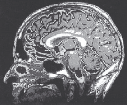
The special advantage of MRI is that it can image soft tissues (Fig. I14.2), whereas X-rays are largely used for imaging hard, bony structures and abnormally dense regions, such as tumours. In fact, the invisibility of hard structures in MRI is an advantage, as it allows the imaging of structures encased by bone, such as the brain and the spinal cord. X-rays are known to be dangerous on account of the ionization they cause; the high magnetic fields used in MRI may also be dangerous but, apart from anecdotes about the extraction of loose fillings from teeth, there is no convincing evidence of their harmfulness, and the technique is considered safe.

Fig. I14.2 The great advantage of MRI is that it can display soft tissue, such as in this cross-section through a patient’s head. (Courtesy of the University of Manitoba.)
* For technical reasons, it is more common to vary the magnitude of the phase gradient.