The Brain

When you think about your brain, you’re thinking with your brain—sending billions of neurotransmitter molecules across countless millions of synapses. Indeed, say neuroscientists, “the mind is what the brain does” (Minsky, 1986).
Even in a permanently motionless body, the brain—and mind—may, in some cases, be active. One hospitalized 23-year-old woman showed no outward signs of conscious awareness after being in a traffic accident. Nevertheless, when researchers asked her to imagine playing tennis or moving around her home, brain scans revealed activity like that of healthy volunteers (Owen et al., 2006). As she imagined playing tennis, for example, an area of her brain controlling arm and leg movements became active.
“I am a brain, Watson. The rest of me is a mere appendix.”
Sherlock Holmes, in Arthur Conan Doyle’s “The Adventure of the Mazarin Stone”
Close-Up: Tools of Discovery, describes some tools scientists use to explore the brain-mind connection. For centuries, we had no device high-powered yet gentle enough to reveal a living brain’s activity. Now we are living in the golden age of brain science, moving closer and closer to understanding where and how the mind’s functions are tied to the brain. To be learning about the brain now is like studying geography while the early explorers were mapping the world. Let’s begin our own exploration of the brain.
Older Brain Structures

Brain structures determine our abilities. In sharks and other primitive vertebrates (animals with backbones), a not-so-complex brain mainly handles basic survival functions: breathing, resting, and feeding. In lower mammals, such as rodents, a more complex brain enables emotion and greater memory. In advanced mammals, such as humans, a brain that processes more information also enables the ability to plan ahead.
This increasing complexity arises from new brain systems built on top of the old, much as new layers cover old ones in the Earth’s landscape. Digging down, one discovers the fossil remnants of the past—brainstem components performing for us much as they did for our distant ancestors. Let’s start with the brain’s basement and work up.
Tools of Discovery—Having Our Head Examined
C L O S E - U P
2-7 What are some techniques for studying the brain?
In the past, brain injuries provided clues to brain-mind connections. For example, damage to one side of the brain often caused paralysis on the body’s opposite side. Noting this, physicians guessed that the body’s right side is wired to the brain’s left side, and vice versa. Others linked vision problems with damage to the back of the brain, and speech problems with damage to the left-front brain. Gradually, a map of the brain began to emerge.
Now a new generation of mapmakers is at work charting formerly unknown territory, stimulating various brain parts and watching the results. Some use microelectrodes to snoop on the messages of individual neurons. Some attach larger electrodes to the scalp to eavesdrop with an EEG (electroencephalograph) on the chatter of billions of neurons. Others use scans that peer into the thinking, feeling brain and give us a Superman-like ability to see what’s happening.
The PET (positron emission tomography) scan tracks the brain’s favorite food, the sugar glucose. Knowing that active neurons are glucose hogs, researchers give the person a form of temporarily radioactive glucose. The PET scan then detects where this “food for thought” goes by locating the radioactivity. Rather like weather radar showing rain activity, PET-scan “hot spots” show which brain areas are most active as the person solves math problems, looks at images of faces, or daydreams.
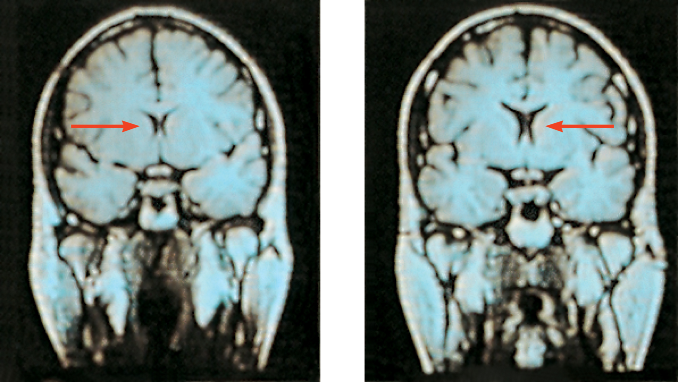
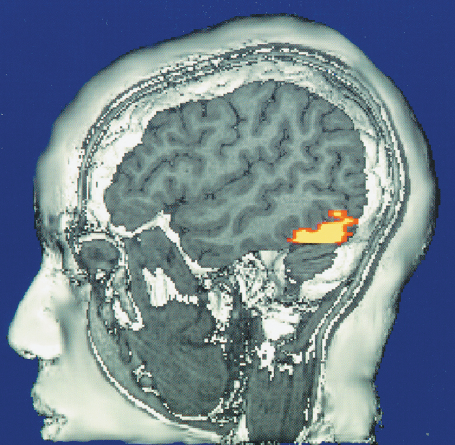
MRI (magnetic resonance imaging) scans capture images of brain structures by briefly disrupting activity in brain molecules. Researchers first position the person’s head in a strong magnetic field, which aligns the spinning atoms of brain molecules. Then, with a brief pulse of radio waves, they disrupt the spinning. When the atoms return to their normal spin, they give off signals that provide a detailed picture of soft tissues, including the brain. MRI scans have revealed, for example, that some people with schizophrenia, a disabling psychological disorder, have enlarged fluid-filled brain areas (Figure 2.6).
RETRIEVE + REMEMBER
Question 2.10
Match the type of neuron to its description.
| Technique |
|
| Description |
|
1. b, 2. a, 3. c
A special application of MRI, fMRI (functional MRI), also reveals the brain’s functions. Where the brain is especially active, blood goes. By comparing MRI scans taken less than a second apart, researchers can watch parts of the brain activate as a person thinks or acts in certain ways. As the person looks at a photo, for example, the fMRI shows blood rushing to the back of the brain, which processes visual information (see Figure 2.17). The technology enables a very crude sort of mind reading, as some neuroscientists showed. They scanned 129 people’s brains as they did eight different mental tasks (such as reading, gambling, and rhyming). Later, viewing another person’s brain images, they were able, with 80 percent accuracy, to predict which of these mental tasks the person was doing (Poldrack et al., 2009).
What the telescope did for astronomy, these brain-snooping tools are doing for psychology. By revealing how the living, working brain divides its labor, these tools have taught us more about the brain in the past 30 years than we had learned in the prior 30,000 years. 
The Brainstem
2-8 What are the functions of the brainstem and its related structures?
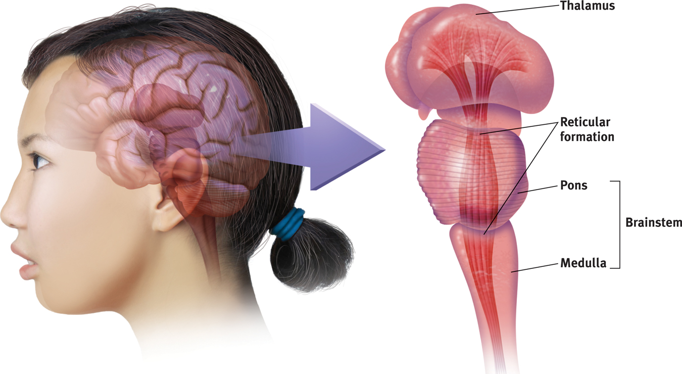
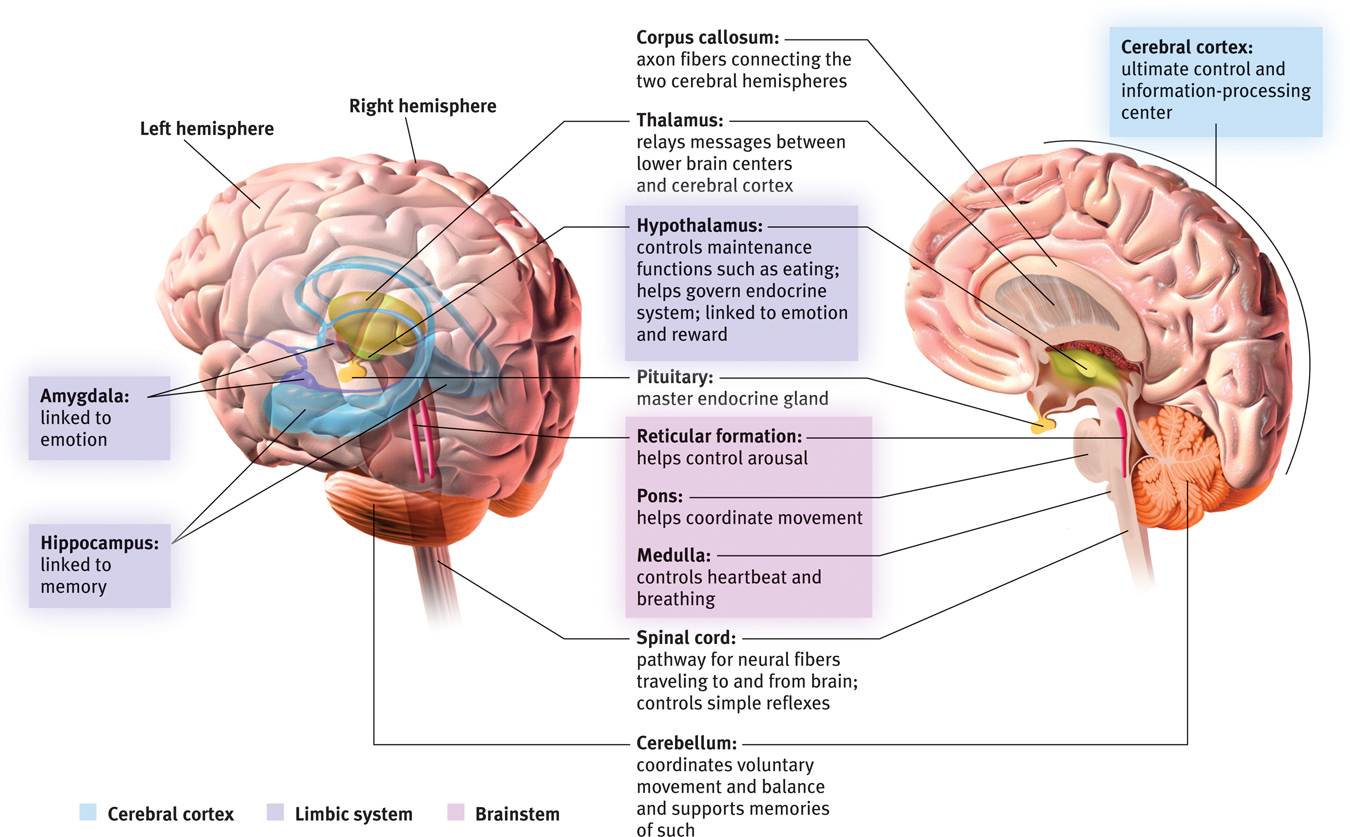
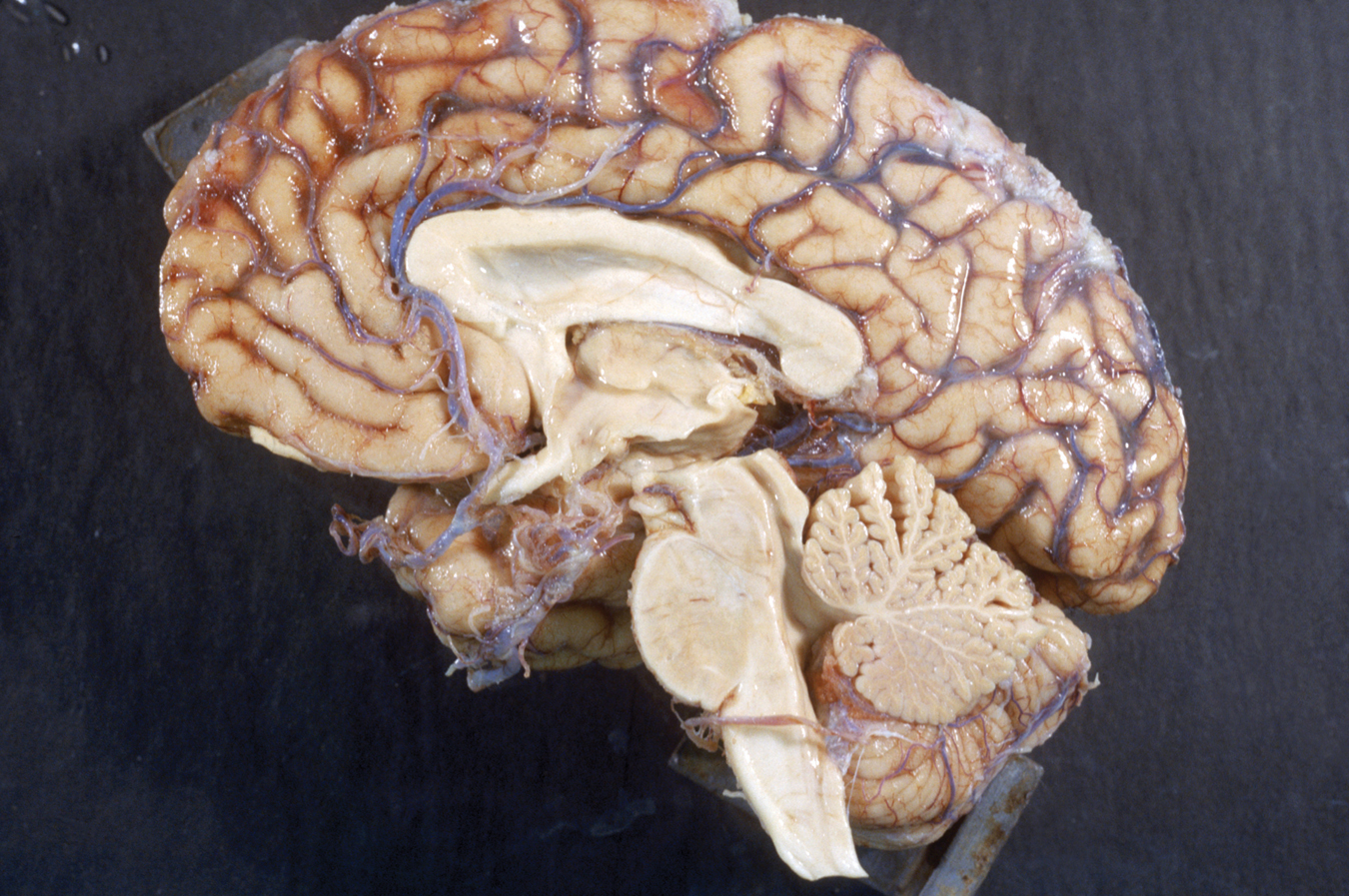
The brain’s oldest and innermost region is the brainstem. It begins where the spinal cord swells slightly after entering the skull. This slight swelling is the medulla (Figure 2.7). Here lie the controls for your heartbeat and breathing. As some severely brain-damaged patients illustrate, no higher brain or conscious mind is needed to orchestrate our heart’s pumping and lungs’ breathing. The brainstem handles those tasks.
38
39
Just above the medulla sits the pons, a brainstem area that helps coordinate movements. If a cat’s brainstem is severed from the rest of the brain above it, the animal will still breathe. It will even run, climb, and groom (Klemm, 1990). But cut off from the brain’s higher regions, it won’t purposefully run or climb to get food.
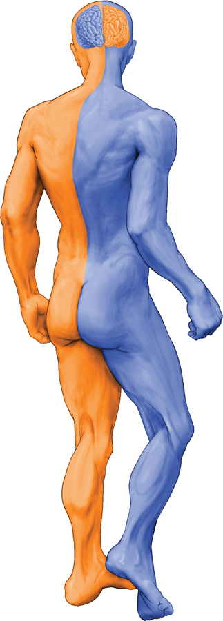
The brainstem is a crossover point. Here, you’ll find a peculiar sort of cross-wiring, with most nerves to and from each side of the brain connecting to the body’s opposite side. Thus, the right brain controls the left side of the body, and vice versa (Figure 2.8). This cross-wiring is one of the brain’s many surprises.
RETRIEVE + REMEMBER
Question 2.11
Nerves from the left side of the brain are mostly linked to the ________ side of the body, and vice versa.
right
The Thalamus
Sitting at the top of the brainstem is the thalamus. This joined pair of egg-shaped structures acts as the brain’s sensory control center. The thalamus receives information from all your senses except smell, and it forwards the messages to brain regions that deal with seeing, hearing, tasting, and touching. Messages flow through this hub on their way to their final destination. In addition to incoming sensory messages, your thalamus receives replies from some higher brain regions. It forwards these replies to other brain areas (your medulla and cerebellum) for processing.
The Reticular Formation
Inside the brainstem, between your ears, lies your reticular (“netlike”) formation . This neuron network extends upward from your spinal cord, through your brainstem, and into your thalamus (see Figure 2.7). This long structure acts as a filter for some of the sensory messages traveling from your spinal cord to your thalamus, relaying important information to other brain areas, and controlling arousal.
In 1949, researchers discovered that electrically stimulating the reticular formation of a sleeping cat almost instantly produced an awake, alert animal (Moruzzi & Magoun, 1949). When a cat’s reticular formation was cut off from higher brain regions, without damaging the nearby sensory pathways, the effect was equally dramatic. The cat lapsed into a coma and never awakened. The conclusion? The reticular formation enables arousal.
40
The Cerebellum
At the rear of the brainstem is the cerebellum, meaning “little brain,” which is what its two wrinkled halves resemble (Figure 2.9). This baseball-sized structure plays an important role in a lot that happens just outside your awareness. Quickly answer these questions. How much time has passed since you woke up this morning? Does your chair feel different from the back of your hand? How’s your mood? If you answered those questions easily, thank your cerebellum.
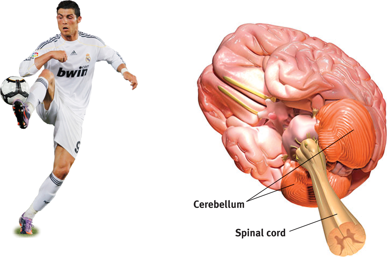
Your cerebellum helps you judge time, discriminate sounds and textures, and control your emotions (Bower & Parsons, 2003). It also coordinates voluntary movement. When soccer player Ronaldo masterfully controls the ball, give his cerebellum some credit. If you injured your cerebellum or drugged it with alcohol, you would have trouble walking, keeping your balance, or shaking hands. The cerebellum also helps process and store memories for things we cannot consciously recall, such as how we ride a bicycle. (Stay tuned for more about this in Chapter 7.)
· · ·
Note: These older brain functions all occur without any conscious effort. Once again, we see one of our Big Ideas at work: Our two-track brain processes most information outside of our awareness. We are aware of the results of our brain’s labor (say, our current visual experience) but not of how we construct the visual image. Likewise, whether we are asleep or awake, our brainstem manages its life-sustaining functions, freeing our newer brain regions to think, talk, dream, or savor a memory.
RETRIEVE + REMEMBER
Question 2.12
In what brain region would damage be most likely to (1) disrupt your ability to skip rope? (2) disrupt your ability to hear and taste? (3) perhaps leave you in a coma? (4) cut off the very breath and heartbeat of life?
1. cerebellum, 2. thalamus, 3. reticular formation, 4. medulla
The Limbic System
2-9 What are the structures and functions of the limbic system?
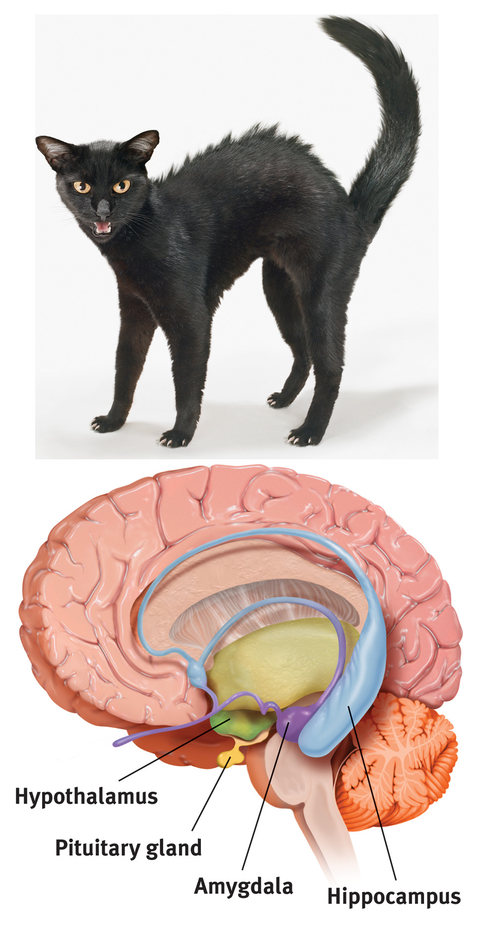
We’ve traveled through the brain’s oldest parts, but we’ve not yet reached its newest and highest regions, the cerebral hemispheres (the two halves of the brain). Between the oldest and newest brain areas lies the limbic system (limbus means “border”). The limbic system contains the amygdala, the hypothalamus, and the hippocampus (Figure 2.10). The hippocampus processes conscious memories. Animals or humans who lose their hippocampus to surgery or injury also lose their ability to form new memories of facts and events. Chapter 7 explains how our two-track mind processes our memories. For now, let’s look at the limbic system’s links to emotions such as fear and anger, and to basic motives such as those for food and sex.
41
THE AMYGDALA The amygdala—two lima-bean-sized neural clusters —enable aggression and fear. In 1939, researchers surgically removed a rhesus monkey’s amygdala, turning the normally ill-tempered animal into the most mellow of creatures (Klüver & Bucy, 1939).
What, then, might happen if we electrically stimulated the amygdala of a normally mellow domestic animal, such as a cat? Do so in one spot and the cat prepares to attack, hissing with its back arched, its pupils wide, its hair on end (see Figure 2.10). Move the electrode only slightly within the amygdala, cage the cat with a small mouse, and now it cowers in terror.
These experiments have confirmed the amygdala’s role in emotions, including the processing of emotional memories and the perception of rage and fear. Consider a woman whose amygdala was destroyed by a rare genetic disease. Whether facing a snake, doing public speaking, or being threatened with a gun, she experiences no fear (Feinstein et al., 2010).
A critical thinker should be careful here. The brain is not neatly organized into structures that reflect our types of behaviors and feelings. When we feel or act in aggressive and fearful ways, there is neural activity in all levels of our brain, not just in the amygdala. If you charge a car’s dead battery, you can activate the engine. Yet the battery is merely one link in the whole working system.
RETRIEVE + REMEMBER
Question 2.13
Electrical stimulation of a cat’s amygdala provokes angry reactions, suggesting the amygdala’s role in aggression. Which ANS division is activated by such stimulation?
The sympathetic nervous system
THE HYPOTHALAMUS Just below (hypo) your thalamus is your hypothalamus, an important link in the chain of command for bodily maintenance. Some neural clusters in the hypothalamus influence hunger. Others regulate thirst, body temperature, and sexual behavior. Together, they help you maintain a steady internal state.
As the hypothalamus monitors your body state, it tunes in to your blood chemistry and any incoming orders from other brain parts. For example, picking up signals from your brain’s information-processing center, the cerebral cortex, that you are thinking about sex, your hypothalamus will secrete hormones. These hormones will in turn trigger the nearby gland of the endocrine system, your pituitary (see Figure 2.10), to influence your sex glands to release their hormones. These will intensify the thoughts of sex in your cerebral cortex. Once again, we see the interplay between the nervous and endocrine systems: The brain influences the endocrine system, which in turn influences the brain.)
A remarkable discovery about the hypothalamus illustrates how progress in science often occurs—when curious, smart-thinking investigators keep an open mind. Two young psychologists, James Olds and Peter Milner (1954), were trying to implant electrodes in rats’ reticular formations when they made a magnificent mistake. In one rat, they placed the electrode incorrectly. Curiously, the rat kept returning to the location where it had been stimulated by this misplaced electrode, as if seeking more stimulation. When Olds and Milner discovered that they had actually placed the device in a region of the hypothalamus, they realized they had stumbled upon a brain center that provides pleasurable rewards (Olds, 1975).
In later studies, when rats were allowed to control their own stimulation in these and other areas, they did so at a feverish pace—pressing a pedal up to 7000 times an hour, until they dropped from exhaustion. Moreover, to get this stimulation, they would even cross an electrified floor that a starving rat would not cross to reach food (Figure 2.11).

Similar reward centers in or near the hypothalamus were later discovered in many other species, including goldfish, dolphins, and monkeys. In fact, animal research has revealed both a general reward system that triggers the release of the neurotransmitter dopamine, and specific centers associated with the pleasures of eating, drinking, and sex. Animals, it seems, come equipped with built-in systems that reward activities essential to survival.
Do you and I have limbic centers for pleasure? Indeed we do. When hearing or anticipating music that gives us great pleasure, our brain releases large amounts of dopamine (Salimpoor et al., 2011). To calm violent patients, one neurosurgeon implanted electrodes in such limbic system areas. Stimulated patients reported mild pleasure. Unlike Olds’ rats, however, they were not driven to a frenzy (Deutsch, 1972; Hooper & Teresi, 1986).
· · ·
42
We’ve finished our tour of the older brain structures. Figure 2.12 will help you place the key brain areas we’ve discussed, as well as the cerebral cortex, our next topic.
RETRIEVE + REMEMBER
Question 2.14
What are the three key structures of the limbic system, and what functions do they serve?
(1) The amygdala is involved in aggression and fear responses. (2) The hypothalamus is involved in bodily maintenance, pleasurable rewards, and control of the hormonal systems. (3) The hippocampus processes memory.
The Cerebral Cortex
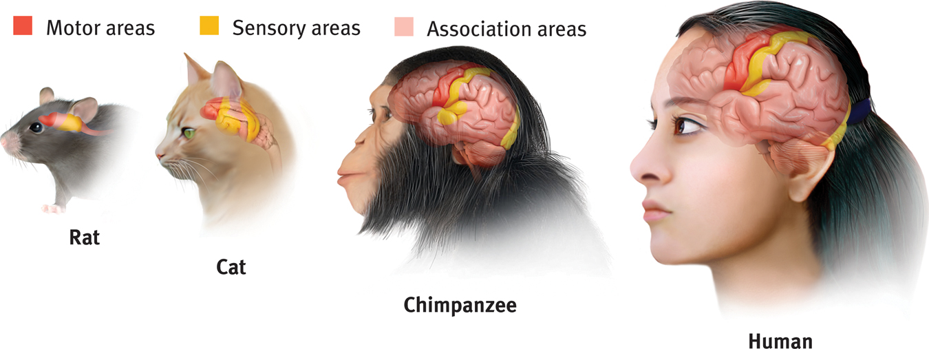
Older brain networks sustain basic life functions and enable memory, emotions, and basic drives. High above these older structures are the two large cerebral hemispheres, which contribute 85 percent of the brain’s weight. Covering those hemispheres, like bark on a tree, is the cerebral cortex, a thin surface layer of interconnected neurons. Its newer neural networks form specialized work teams that enable your thinking, sensing, and speaking. The cerebral cortex is your brain’s thinking crown, your body’s ultimate control and information-processing center.
Structure of the Cortex
2-10 What are the four lobes of the cerebral cortex, and where are they located?
If you opened a human skull, exposing the brain, you would see a wrinkled organ, shaped somewhat like the meat of an oversized walnut. Without these wrinkles, a flattened cerebral cortex would require triple the area—roughly that of a very large pizza. The brain’s left and right hemispheres are filled mainly with axons connecting the cortex to the brain’s other regions. The cerebral cortex—that thin surface layer—contains some 20 to 23 billion nerve cells and 300 trillion synaptic connections (de Courten-Myers, 2005). Being human takes a lot of nerve.
43
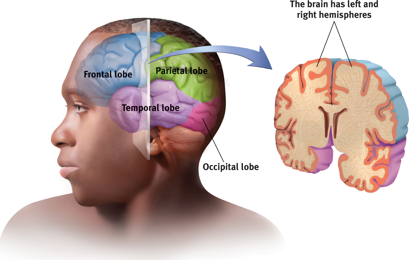
Each hemisphere’s cortex is subdivided into four lobes, separated by deep folds (Figure 2.13). You can roughly trace the four lobes, starting with both hands on your forehead. The frontal lobes lie directly behind your forehead. As you move your hands over the top of your head, toward the rear, you’re sliding over your parietal lobes. Continuing to move down, toward the back of your head, you’ll slide over your occipital lobes. Now move each hand forward, to the sides of your head, and just above each ear you’ll find your temporal lobes. Each hemisphere has these four lobes. Each lobe carries out many functions. And many functions require the cooperation of several lobes.
Functions of the Cortex
2-11 What are the functions of the motor cortex, somatosensory cortex, and association areas?
More than a century ago, surgeons found damaged areas of the cerebral cortex during autopsies of people who had been partially paralyzed or speechless. This rather crude evidence was interesting, but it did not prove that specific parts of the cortex control complex functions like movement or speech. After all, if the entire cortex controlled speech and movement, damage to almost any area might produce the same effect. A TV with a cut power cord would go dead, but we would be fooling ourselves if we thought we had “localized” the picture in the cord.
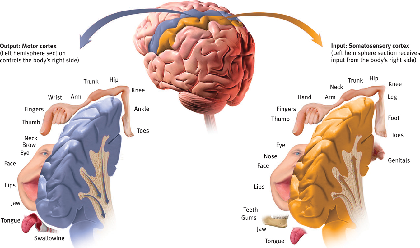
MOTOR FUNCTIONS Early scientists had better luck showing simpler brain-behavior links. In 1870, for example, German physicians Gustav Fritsch and Eduard Hitzig made an important discovery. By electrically stimulating parts of a dog’s cortex, they could make other parts of its body move. The movement happened only when they stimulated an arch-shaped region at the back of the dog’s frontal lobe, running roughly ear-to-ear across the top of the brain. Moreover, if they stimulated this region in the left hemisphere, the dog’s right leg would move. And if they stimulated part of the right hemisphere, the opposite leg—on the left—reacted. Fritsch and Hitzig had discovered what is now called the motor cortex.
Lucky for brain surgeons and their patients, the brain has no sensory receptors. Knowing this, Otfrid Foerster and Wilder Penfield were able to map the motor cortex in hundreds of wide-awake patients by stimulating different cortical areas and observing the body’s responses. They discovered that body areas requiring precise control, such as the fingers and mouth, occupied the greatest amount of cortical space (Figure 2.14).
44
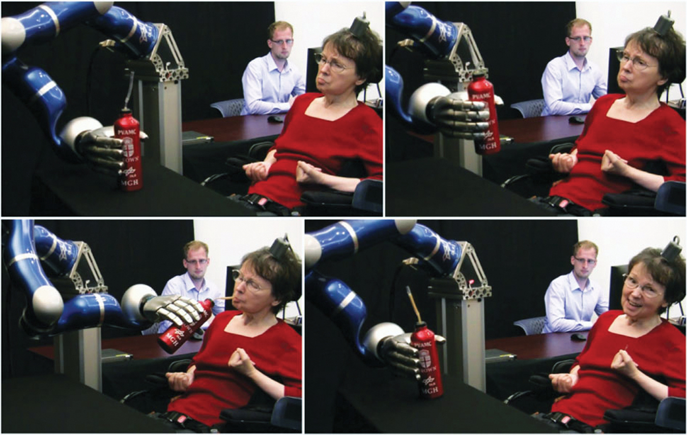
As is so often the case in science, new answers triggered new questions. We now know that electrically stimulating the motor cortex can cause body parts to move. What might happen, some researchers are asking, if we implanted a device to detect motor cortex activity? Could brain-controlled computer devices direct a robotic limb to move in soldiers who have lost arms or legs during combat? Could they help severely paralyzed people learn to command a cursor to send texts or e-mails or work online? Clinical trials are now under way with people who have suffered paralysis or amputation (Andersen et al., 2010; Nurmikko et al., 2010) (Figure 2.15).
45
RETRIEVE + REMEMBER
Question 2.15
Try moving your right hand in a circular motion, as if polishing a car. Then start your right foot doing the same motion as your hand. Now reverse the right foot’s motion, but not the hand’s. Finally, try moving the left foot opposite to the right hand.
|
1. The right limbs’ activities interfere with each other because both are controlled by the same (left) side of your brain. 2. Opposite sides of your brain control your left and right limbs, so the reversed motion causes less interference.
SENSORY FUNCTIONS The motor cortex sends messages out to the body. What parts of the cortex receive incoming messages from our senses of touch and movement? Penfield supplied the answer: the somatosensory cortex, which runs parallel to the motor cortex and just behind it at the front of the parietal lobes (see Figure 2.14). Stimulate a point on the top of this band of tissue, and a person reports being touched on the shoulder. Stimulate some point on the side, and the person feels something on the face.
The more sensitive a body region, the larger the somatosensory cortex area devoted to it. Why do we kiss with our lips rather than rub elbows? Our supersensitive lips project to a larger brain area than do our arms (see Figure 2.14). Similarly, rats have a large brain area devoted to their whisker sensations, and owls to their hearing sensations.
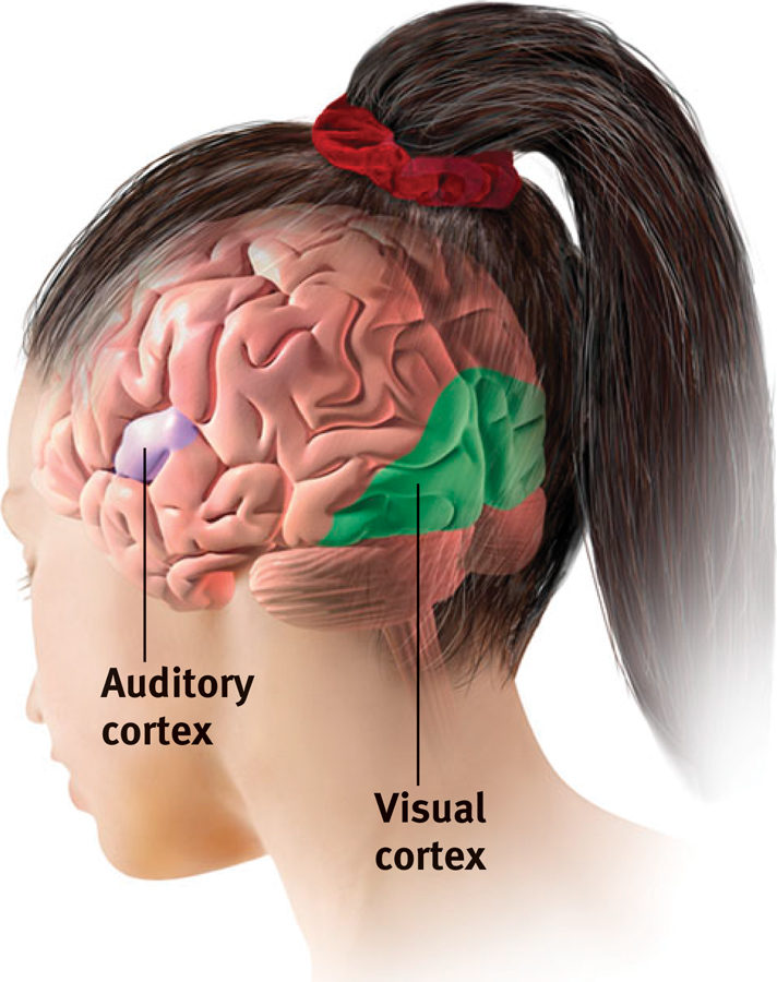
Your somatosensory cortex is a very powerful tool for processing information from your skin senses and from movements of your body parts. But it isn’t the only area of the cortex that receives input from your senses. If you have normal vision, you are at this moment receiving visual information in the visual cortex in your occipital lobes, at the back of your brain (Figure 2.16). A friend of mine, who lost much of his right occipital lobe when a tumor was removed, became blind to the left half of his field of vision. Stimulated in your occipital lobes, you might see flashes of light or dashes of color. (In a sense, we do have eyes in the back of our head!) From your occipital lobes, visual information goes to other areas that specialize in tasks such as identifying words, detecting emotions, and recognizing faces (Figure 2.17).
Any sound you now hear is processed by your auditory cortex in your temporal lobes (just above your ears; see Figure 2.16). Most of this auditory information travels a roundabout route from one ear to the auditory receiving area above your opposite ear. If stimulated in your auditory cortex, you alone might hear a sound. People with schizophrenia sometimes have auditory hallucinations (false sensory experiences). MRI scans taken during these hallucinations show active auditory areas in the temporal lobes (Lennox et al., 1999).
RETRIEVE + REMEMBER
Question 2.16
Our brain’s_________ cortex registers and processes body touch and movement sensations. The ___________cortex controls our voluntary movements.
somatosensory; motor
ASSOCIATION AREAS So far, we have pointed out small areas of the cortex that receive messages from our senses, and other small areas that send messages to our muscles. Together, these areas occupy about one-fourth of the human brain’s thin, wrinkled cover. What, then, goes on in the vast remaining regions of the cortex? In these association areas (peach colored in Figure 2.18), neurons are busy with higher mental functions—many of the tasks that make us human.
46
Electrically probing an association area won’t trigger any observable response. So, unlike the sensory and motor areas, association area functions can’t be neatly mapped. Their silence has led to what Donald McBurney (1996, p. 44) called “one of the hardiest weeds in the garden of psychology”—the claim that we really use only 10 percent of our brain. (Time for some critical thinking: Wouldn’t that mean that there is a 90 percent chance that a bullet to your brain would land in an unused area?) Brain-damaged animals and humans provide evidence that association areas are not unused. Rather, these brain areas interpret, integrate, and act on sensory information and link it with stored memories—a very important part of thinking.
Association areas are found in all four lobes. In the frontal lobes, they enable judgment, planning, and processing of new memories. People with damaged frontal lobes may have intact memories, high intelligence scores, and great cake-baking skills. Yet they would not be able to plan ahead to begin baking a cake for a loved one’s birthday (Huey et al., 2006).
Frontal lobe damage can also alter personality and remove a person’s inhibitions. Consider the classic case of railroad worker Phineas Gage. One afternoon in 1848, Gage, then 25 years old, was using an iron rod to pack gunpowder into a rock. A spark ignited the gunpowder, shooting the rod up through his left cheek and out the top of his skull, causing massive damage to his frontal lobes (Figure 2.19a). To everyone’s amazement, he was immediately able to sit up and speak, and after the wound healed, he returned to work. But the friendly, soft-spoken Phineas Gage was now irritable, profane, and dishonest. This person, said his friends, was “no longer Gage.” His mental abilities and memories were intact, but his personality was not. (Although Gage lost his railroad job, he did, over time, adapt to his injury and find work as a stagecoach driver [Macmillan & Lena, 2010].)

With his frontal lobes ruptured, Gage’s moral compass had disconnected from his behavior. More recent studies of people with damaged frontal lobes have revealed similar losses. Without the frontal lobe brakes on their impulses they, too, became less inhibited. Moreover, their moral judgments seem untouched by normal emotions. Would you agree with the idea of pushing someone in front of a runaway train to save five others? Most people do not, but those with damage to a brain area behind the eyes often do (Koenigs et al., 2007).
Damage to association areas in other lobes would result in different losses. If a stroke or head injury destroyed part of your parietal lobes, you might lose mathematical and spatial reasoning. If the damaged area was on the underside of the right temporal lobe, which lets you recognize faces, you would still be able to describe facial features and to recognize someone’s gender and approximate age. Yet you would be strangely unable to identify the person as, say, Lady Gaga or even your grandmother.
47
Nevertheless, complex mental functions don’t reside in any one place. There is no one spot in a rat’s small association cortex that, when damaged, will wipe out its ability to learn or remember a maze. And as we’ll see in Chapter 8, distinct neural networks in the human brain work together to enable language. Memory, language, and attention are the products of interaction among distinct brain areas (Knight, 2007). Ditto for religious experience. More than 40 brain regions become active in different religious states, such as prayer and meditation, indicating that there is no simple “God spot” (Fingelkurts & Fingelkurts, 2009). The point to remember: Our mental experiences arise from coordinated brain activity.
RETRIEVE + REMEMBER
Question 2.17
Why are association areas important?
Association areas are involved in higher mental functions—interpreting, integrating, and acting on information processed in other areas.
The Brain’s Plasticity
2-12 When damaged, can the brain repair or reorganize itself?

Our brains are sculpted not only by our genes but also by our experiences. In Chapter 3, we’ll focus more on how experience molds the brain. For now, let’s turn to another aspect of the brain’s plasticity: its ability to modify itself after damage.
Some brain-damage effects described earlier can be traced to two hard facts:
- Severed neurons, unlike cut skin, usually do not repair themselves. (If your spinal cord were severed, you probably would be permanently paralyzed.)
- Some brain functions seem forever linked to specific areas. One newborn who suffered damage to the facial recognition areas on both temporal lobes later remained unable to recognize faces (Farah et al., 2000).
But there is good news: Thanks to the brain’s impressive plasticity, some brain tissue can reorganize in response to damage. Under the surface of our awareness, the brain is constantly changing, building new pathways as it adjusts to little mishaps and new experiences.
On a larger, more dramatic scale, plasticity sometimes occurs after serious damage, especially in young children (Kolb, 1989). If a slow-growing left-hemisphere tumor disrupts language, the right hemisphere may take over the task (Thiel et al., 2006). If a finger is lost, the somatosensory cortex that received its input will begin to pick up signals from the neighboring fingers, which then become more sensitive (Fox, 1984). Blindness or deafness makes unused brain areas available for other uses (Amedi et al., 2005). This plasticity helps explain why deaf people who learned sign language before any other may have better-than-average peripheral vision (Bosworth & Dobkins, 1999). Without stimulation from sounds, a temporal lobe area normally dedicated to hearing is free to process other signals, such as those from the visual system.
Although the brain often attempts self-repair by reorganizing existing tissue, it sometimes tries to mend itself by producing new neurons. This process, known as neurogenesis, has been found in adult mice, birds, monkeys, and humans (Jessberger et al., 2008). These baby neurons are born deep in the brain. They may then migrate elsewhere and form connections with neighboring neurons (Aimone et al., 2010; Gould, 2007).
Might new drugs spur the production of new nerve cells? Stay tuned. As you read this sentence, companies are working on such possibilities. In the meantime, we can all benefit from natural aids to neurogenesis, such as exercise, sleep, and nonstressful but stimulating environments (Iso et al., 2007; Pereira et al., 2007; Stranahan et al., 2006).
Our Divided Brain
2-13 What is a split brain, and what does it reveal about the functions of our left and right hemispheres?
“You wouldn’t want to have a date with the right hemisphere.”
Michael Gazzaniga, 2002
Our brain’s look-alike left and right hemispheres serve different functions. This lateralization is clear after some types of brain damage. Language processing seems to reside mostly in your left hemisphere. More than a century’s research has shown that left hemisphere accidents, strokes, and tumors could leave you unable to read, write, speak, do arithmetic, and understand others. Similar right hemisphere damage seldom has such dramatic effects.
48
Does this mean that the right hemisphere is just along for the ride—a silent junior partner or “minor” hemisphere? Many believed this was the case until 1960, when researchers found that the “minor” right hemisphere was not so limited after all. The unfolding of this discovery is another one of psychology’s fascinating stories.
Splitting the Brain: One Skull, Two Minds
In 1961, two neurosurgeons believed that the uncontrollable seizures of some patients with severe epilepsy could be caused by abnormal brain activity bouncing back and forth between the two cerebral hemispheres. If so, they wondered, could they put an end to this biological tennis game by cutting through the corpus callosum, the wide band of axon fibers connecting the two hemispheres and carrying messages between them (Figure 2.20)? The neurosurgeons knew that psychologists Roger Sperry, Ronald Myers, and Michael Gazzaniga had divided cats’ and monkeys’ brains in this manner, with no serious ill effects.
So the surgeons operated. The result? The seizures all but disappeared. The patients with these split brains were surprisingly normal, their personality and intellect hardly affected. Waking from surgery, one even joked that he had a “splitting headache” (Gazzaniga, 1967). By sharing their experiences with us, these patients have greatly expanded our understanding of interactions between the intact brain’s two hemispheres.
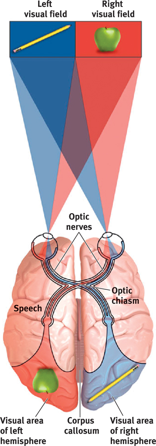
To appreciate these studies, we need to focus for a minute on the peculiar nature of our visual wiring, illustrated in Figure 2.21. Note that each eye receives sensory information from the entire visual field. In each eye, information from the left half of your field of vision goes to your right hemisphere, and information from the right half of your visual field goes to your left hemisphere, which usually controls speech. In an intact brain, data received by either hemisphere are quickly transmitted to the other side, across the corpus callosum. In a person with a severed corpus callosum, this information sharing does not take place.
Knowing these facts, Sperry and Gazzaniga could send information to a patient’s left hemisphere by having the person stare at a dot and by then flashing a stimulus (a word or photo) to the right of the dot. To send a message to the right hemisphere, they would flash the item to the left of the dot.
They could do this with you, too, but in your intact brain the hemisphere receiving the information would instantly pass the news to the other side. Because the split-brain surgery had cut the communication lines between the hemispheres, the researchers could, with these patients, quiz each hemisphere separately.
49
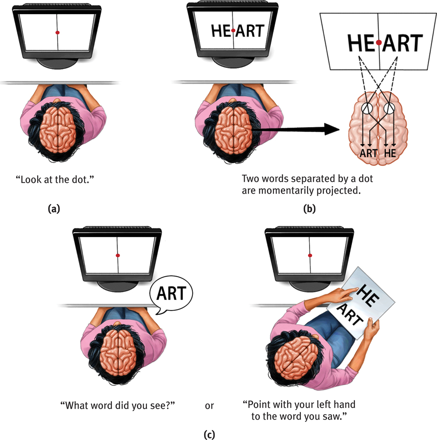
In an early experiment, Gazzaniga (1967) flashed the word HEART across the screen in such a way that HE appeared to the left of the dot, and ART appeared to the right (Figure 2.22b). Asked to say what they saw, the patients reported the letters sent to the left hemisphere—“ART.” Asked to point with their left hand to what they saw, they pointed to the letters sent to the right hemisphere—“HE” (Figure 2.22c). One skull was housing two minds.

A few people who have had split-brain surgery have been bothered for a time by the unruly independence of their left hand. It seemed the left hand truly didn’t know what the right hand was doing. One hand might unbutton a shirt while the other buttoned it, or put grocery store items back on the shelf after the other hand put them in the cart. It was as if each hemisphere was thinking, “I’ve half a mind to wear my green (blue) shirt today.” Indeed, said Sperry (1964), split-brain surgery leaves people “with two separate minds” (Figure 2.23).
Which hemisphere resolves disagreements when the “two minds” are at odds? If a split-brain patient follows an order (“Walk”) sent to the right hemisphere, the left hemisphere won’t know why it did so. But rather than say “I don’t know,” the left hemisphere does a strange thing. It instantly invents—and apparently believes—an explanation (“I’m going into the house to get a Coke”). Thus, Gazzaniga (1989), who has called split-brain patients “the most fascinating people on Earth,” concluded that the conscious left hemisphere is an “interpreter” that instantly constructs theories to explain our behavior. And so it goes for us all. The brain runs largely on autopilot: Often it feels and acts and then explains later (Kahneman, 2011).
RETRIEVE + REMEMBER
Question 2.18
(1) If we flash a red light to the right hemisphere of a person with a split brain, and flash a green light to the left hemisphere, will each observe its own color? (2) Will the person be aware that the colors differ? (3) What will the person verbally report seeing?
1. yes, 2. no, 3. green
50
Right-Left Differences in Intact Brains
So, what about the 99.99+ percent of us with undivided brains? Does each of our hemispheres also perform distinct functions? Several different types of studies indicate they do. When a person performs a perceptual task, for example, brain waves, bloodflow, and glucose consumption reveal increased activity in the right hemisphere. When the person speaks or calculates, activity increases in the left hemisphere.
If you could peek into an operating room at the beginning of some types of brain surgery, you could watch a dramatic demonstration of lateralization. To locate the patient’s language centers, the surgeon injects a sedative into the neck artery feeding blood to the left hemisphere, which usually controls speech. Before the injection, the patient is lying down, arms in the air, chatting with the doctor. Can you predict what probably happens when the drug puts the left hemisphere to sleep? Within seconds, the patient’s right arm falls limp. If the left hemisphere is controlling language, the patient will be speechless until the drug wears off.
To the brain, language is language, whether spoken or signed. Just as hearing people usually use the left hemisphere to process spoken language, deaf people usually use the left hemisphere to process sign language (Corina et al., 1992; Hickok et al., 2001). Thus, a left-hemisphere stroke disrupts a deaf person’s signing, much as it would disrupt a hearing person’s speaking (Corina, 1998). The same brain area is involved in both. (For more on how the brain enables language, see Chapter 8.)
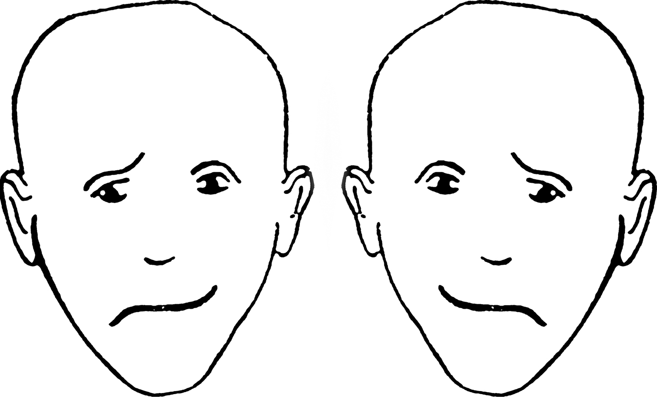
The left hemisphere is good at making quick, exact interpretations of language. But the right hemisphere excels in high-level language processing (Beeman & Chiarello, 1998; Bowden & Beeman, 1998; Mason & Just, 2004). Given an insight problem—“What word goes with boot, summer, and ground?”—the right hemisphere more quickly than the left achieves the solution: camp. As one patient explained after a right-hemisphere stroke, “I understand words, but I’m missing the subtleties.” The right side of the brain is also better than the left at copying drawings, recognizing faces, noticing differences, perceiving emotion, and expressing emotion through the more expressive left side of the face (Figure 2.24). Right-hemisphere damage can greatly disrupt these abilities.
Simply looking at the two hemispheres, so alike to the naked eye, who would suppose they contribute uniquely to the harmony of the whole? Yet a variety of observations—of people with split brains and those with intact brains, and even of other species’ brains—leaves little doubt that we have unified brains with specialized parts (Hopkins & Cantalupo, 2008; MacNeilage et al., 2009).