Memory Storage
7-9 What is the capacity of long-term memory? Are our long-term memories processed and stored in specific locations?
 In Arthur Conan Doyle’s A Study in Scarlet, Sherlock Holmes offers a popular theory of memory capacity:
I consider that a man’s brain originally is like a little empty attic, and you have to stock it with such furniture as you choose…. It is a mistake to think that that little room has elastic walls and can distend to any extent. Depend upon it, there comes a time when for every addition of knowledge you forget something that you knew before.
Contrary to Holmes’ “memory model,” our capacity for storing long-term memories has no real limit. Many endure for a lifetime. Our brains are not like attics, which, once filled, can store more items only if we discard old ones.
Retaining Information in the Brain
I marveled at my aging mother-in-law, a retired pianist and organist. At age 88, her blind eyes could no longer read music. But let her sit at a keyboard and she would flawlessly play any of hundreds of hymns, including ones she had not thought of for 20 years. Where did her brain store those thousands of note patterns?
For a time, some surgeons and memory researchers marveled at what appeared to be vivid memories triggered by stimulating the brain during surgery. Did this prove that our whole past, not just well-practiced music, is “in there,” just waiting to be relived? Further research disproved this idea. The vivid flashbacks were actually new creations of a stressed brain, not real memories (Loftus & Loftus, 1980). We do not store information in single, specific spots, as libraries store their books. As with perception, language, emotion, and much more, memory requires brain networks. Many parts of our brain interact as we encode, store, and retrieve information.
Explicit-Memory System: The Hippocampus and Frontal Lobes
7-10 What roles do the hippocampus and frontal lobes play in memory processing?

Separate brain regions process our explicit and implicit memories. We know this from scans of the brain in action, and from autopsies of people who suffered different types of memory loss.
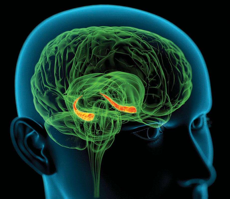
New explicit memories of names, images, and events are laid down via the hippocampus, a limbic system neural center that is our brain’s equivalent of a “save” button (FIGURE 7.6). When brain scans capture the brain forming an explicit memory, they reveal hippocampus activity. Your hippocampus acts as a loading dock where your brain registers and temporarily stores aspects of an event—its smell, feel, sound, and location. Then, like older files shifted to a storeroom, memories migrate for storage elsewhere. This storage process is called memory consolidation.
Your brain’s right and left frontal lobes store different information. Recalling a password and holding it in working memory, for example, would activate your left frontal lobes. Calling up a visual image of last night’s party would more likely activate your right frontal lobe.
A good night’s sleep supports memory consolidation, both in humans and in rats. In experiments, rats have learned the location of a tasty new food. If their hippocampus is removed 3 hours after they locate the food, no long-term memory will form (Tse et al., 2007). If their hippocampus is removed 48 hours later, after doing its work, they still remember that location. During sleep, the hippocampus and brain cortex display rhythmic patterns of activity, as if they were talking to each other (Ji & Wilson, 2007; Mehta, 2007). Researchers suspect that the brain is replaying the day’s experiences as it transfers them to the cortex for long-term storage.
Implicit Memory System: The Cerebellum and Basal Ganglia
7-11 What roles do the cerebellum and basal ganglia play in memory processing?
You could lose your hippocampus and still—thanks to automatic processing—lay down implicit memories of newly conditioned associations and skills (such as golfing). Memory loss following brain damage left one patient unable to recognize her physician as, each day, he shook her hand and introduced himself. One day, after reaching for his hand, she yanked hers back, for the physician had pricked her with a tack in his palm. When he next introduced himself, she refused to shake his hand but couldn’t explain why. Having been classically conditioned, she just wouldn’t do it (LeDoux, 1996).
Your cerebellum, a brain region extending out from the rear of your brainstem, plays an important role in forming and storing memories created by classical conditioning. People with a damaged cerebellum cannot develop some conditioned reflexes. They can’t, for example, link a tone with an oncoming puff of air, so they don’t blink just before the puff, as you and I would learn to do (Daum & Schugens, 1996; Green & Woodruff-Pak, 2000). Implicit memory formation needs the cerebellum.
Your memories of physical skills—walking, cooking, dressing—are also implicit memories. Your basal ganglia, deep brain structures involved in motor movement, help form your memories for these skills (Mishkin, 1982; Mishkin et al., 1997). If you have learned how to ride a bike, thank your basal ganglia.
Although not part of our conscious adult memory system, the reactions and skills we learned during infancy reach far into our future. Can you remember learning to talk and walk as a baby? If you cannot, you are not alone. As adults, our conscious memory of our first three years is blank, an experience called infantile amnesia. To form and store explicit memories, we need a command of language and a well-developed hippocampus. Before age 3, we don’t have those learning tools. 
RETRIEVE + REMEMBER
Question 7.6
 Which parts of the brain are important for implicit memory processing, and which parts play a key role in explicit memory processing?
The cerebellum and basal ganglia are important for implicit memory processing. The frontal lobes and hippocampus are key to explicit memory formation.
Question 7.7
Your friend has experienced brain damage in an accident. He can remember how to tie his shoes but has a hard time remembering anything told to him during a conversation. What’s going on here?
Our explicit conscious memories differ from our implicit memories of skills and classically conditioned responses. The brain areas that process implicit memories apparently escaped damage during the accident.
The Amygdala, Emotions, and Memory
7-12 How do emotions affect our memory processing?
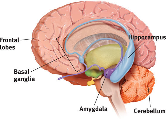
Cerebellum and basal ganglia: implicit memory formation
Amygdala: emotion-related memory formation
Arousal can sear certain events into the brain (Birnbaum et al., 2004; Strange & Dolan, 2004). Excitement or stress (perhaps a time you performed in front of a crowd) triggers your glands to produce stress hormones. By making more glucose energy available to fuel brain activity, stress hormones signal the brain that something important has happened. They also provoke the amygdala (two limbic system, emotion-processing clusters) to boost activity in the brain’s memory-forming areas (Buchanan, 2007; Kensinger, 2007) (FIGURE 7.7).
The resulting emotions often persist without our conscious awareness of what caused them, as one clever experiment demonstrated. The participants were patients with hippocampal damage, which left them unable to form new explicit memories. Researchers first showed them a sad film, and later a happy film. Although the viewers could not consciously recall the films, the sad or happy emotion lingered (Feinstein et al., 2010).
After horrific experiences—a wartime ambush, a house fire, a rape—vivid memories of the event may intrude again and again. The result is “stronger, more reliable memories” (McGaugh, 1994, 2003). The persistence of such memories is adaptive. They alert us to future dangers. By giving us a mental tunnel vision of the remembered event, they reduce our attention to minor details and focus our attention on the central event (Mather & Sutherland, 2012).
Weaker emotions mean weaker memories. In one study, people received a drug that blocked the effects of stress hormones. Later, they had trouble remembering the details of an upsetting story (Cahill, 1994).
“The biology of the mind will be as scientifically important to this [new] century as the biology of the gene [was] to the twentieth century.”
Eric Kandel, acceptance remarks for his 2000 Nobel Prize
Why are some memories so much stronger than others? Emotion-triggered hormonal changes help explain why we long remember exciting or shocking events, such as our first kiss or our whereabouts when learning of a loved one’s death. Psychologists call them flashbulb memories. It’s as if the brain commands, “Capture this!” Ask any adult American where he or she first heard the news of the 9/11 terrorist attacks—that fateful day that the New York Times called “one of those moments in which history splits and we define the world as ‘before and after.’” Five years later, 95 percent of surveyed Americans said they remembered where they were or what they were doing (Pew, 2006). With time, some errors crept in (compared with earlier reports taken right after 9/11). Mostly, however, people’s memories of 9/11 remained consistent over the next two to three years (Conway et al., 2009; Hirst et al., 2009; Kvavilashvili et al., 2009).
Which is more important—your experiences or your memories of them?
Dramatic experiences remain bright and clear in our memory in part because we rehearse them. We think about them and describe them to others. Memories of our best experiences, which we enjoy recalling and recounting, also endure. One study invited 1563 Boston Red Sox and New York Yankees fans to recall the baseball championship games between their two teams in 2003 (Yankees won) and 2004 (Red Sox won). Fans recalled much better the game their team won (Breslin & Safer, 2011).
Synaptic Changes
7-13 How do changes at the synapse level affect our memory processing?
As you read this chapter and learn about memory processes, your brain is changing. Activity in some brain pathways is increasing. Neural network connections are forming and strengthening. Changes are taking place at your synapses—the sites where nerve cells communicate with one another by means of chemical messengers. Experience alters the brain’s neural networks (see Chapter 3).
Eric Kandel and James Schwartz (1982) were able to catch a new memory leaving tracks in neurons of the California sea slug. This simple animal’s nerve cells are unusually large, and researchers have been able to observe how they change during learning. Using electric shocks, they have classically conditioned sea slugs to withdraw their gills when squirted with water, much as we might jump when lightning strikes nearby. By observing the slugs’ neural connections before and after this conditioning, Kandel and Schwartz pinpointed changes. As a slug learns, it releases more of the neurotransmitter serotonin into certain cells. These cells then become more efficient at transmitting signals. Experience and learning can increase—even double—the number of synapses, even in slugs (Kandel, 2012). No wonder the brain area that processes spatial memory grows larger the longer London cab drivers have navigated the maze of streets (Woolett & Maguire, 2011).

As synapses become more efficient, so do neural networks. Sending neurons now release their neurotransmitters more easily. Receiving neurons may grow additional receptor sites. This increased neural efficiency, called long-term potentiation (LTP), enables learning and memory (Lynch, 2002; Whitlock et al., 2006). Several lines of evidence confirm that LTP is a physical basis for memory.
- Rats given a drug that enhanced synaptic efficiency (LTP) learned to run a maze with half the usual number of mistakes (Service, 1994).
- Drugs that block LTP interfere with learning (Lynch & Staubli, 1991).
- Mice that could not produce an enzyme needed for LTP could not learn their way out of a maze (Silva et al., 1992).
After LTP has occurred, an electric current passing through the brain won’t erase old memories. Before LTP, the same current can wipe out very recent memories. This often happens when severely depressed people receive electroconvulsive therapy (Chapter 14). Sports concussions can also wipe out recent memories. Football players and boxers knocked unconscious typically have no memory of events just before the blow to the head (Yarnell & Lynch, 1970). Their working memory had no time to process the information into long-term memory before the shutdown.
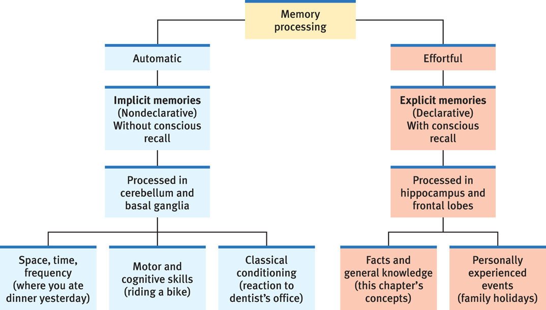
FIGURE 7.8 summarizes the brain’s two-track memory processing and storage system for implicit (automatic) and explicit (effortful) memories.
RETRIEVE + REMEMBER
Question 7.8
Which brain area responds to stress hormones by helping to create stronger memories?
the amygdala
Question 7.9
The neural basis for learning and memory, found at the synapses in the brain’s memory connections, results from brief, rapid stimulation. It is called _______ _______.
long-term potentiation