2.3 The Nervous System
LOQ 2-
nervous system the body’s speedy, electrochemical communication network, consisting of all the nerve cells of the central and peripheral nervous systems.
central nervous system (CNS) the brain and spinal cord.
peripheral nervous system (PNS) the sensory and motor neurons connecting the central nervous system to the rest of the body.
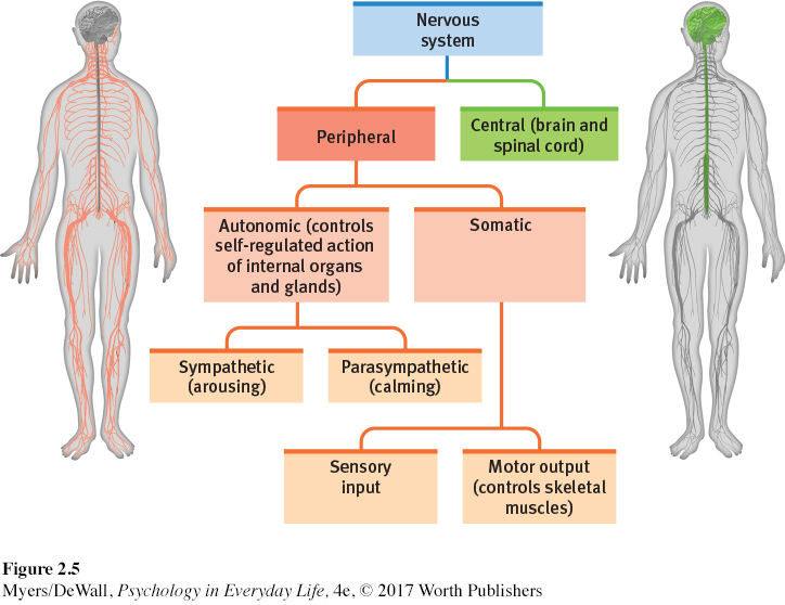
To live is to take in information from the world and the body’s tissues, to make decisions, and to send back information and orders to the body’s tissues. All this happens thanks to your body’s nervous system (FIGURE 2.5). Your brain and spinal cord form the central nervous system (CNS), your body’s decision maker. Your peripheral nervous system (PNS) gathers information from other body parts and transmits CNS decisions to the rest of your body.
nerves bundled axons that form neural cables connecting the central nervous system with muscles, glands, and sense organs.
Nerves are electrical cables formed from bundles of axons. They link your central nervous system with your body’s sensory receptors, muscles, and glands. Your optic nerve, for example, bundles a million axons into a single cable carrying messages from each eye to your brain (Mason & Kandel, 1991). Information travels in your nervous system through three types of neurons.
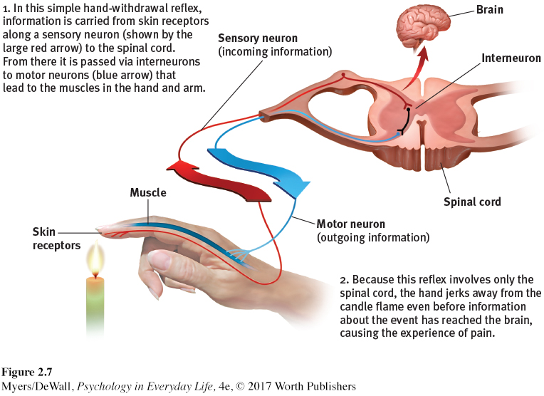
sensory neuron neuron that carries incoming information from the body’s tissues and sensory receptors to the brain and spinal cord.
Sensory neurons carry messages from your body’s tissues and sensory receptors inward to your spinal cord and brain for processing.
motor neuron neuron that carries outgoing information from the brain and spinal cord to the muscles and glands.
Motor neurons carry instructions from your central nervous system outward to your body’s muscles and glands.
interneuron neurons within the brain and spinal cord; communicate internally and process information between sensory inputs and motor outputs.
Interneurons within your brain and spinal cord communicate with one another and process information between the sensory input and motor output.
Your complexity resides mostly in your interneuron systems. Your nervous system has a few million sensory neurons, a few million motor neurons, and billions and billions of interneurons.
The Peripheral Nervous System
somatic nervous system peripheral nervous system division that controls the body’s skeletal muscles. Also called the skeletal nervous system.
autonomic [aw-
The peripheral nervous system has two parts—
sympathetic nervous system autonomic nervous system subdivision that arouses the body, mobilizing its energy.
parasympathetic nervous system autonomic nervous system subdivision that calms the body, conserving its energy.
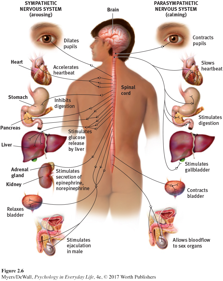
Within your autonomic nervous system, two subdivisions help you cope with challenges (FIGURE 2.6). If something alarms or challenges you (perhaps giving a speech), your sympathetic nervous system will arouse you, making you more alert, energetic, and ready for action. It will increase your heartbeat, blood pressure, and blood-
I [DM] recently experienced my ANS in action. Before sending me into an MRI (magnetic resonance imaging) machine for a shoulder scan, the technician asked if I had ever had claustrophobia (panic feelings when confined). “No, I’m fine,” I assured her, with perhaps a hint of macho swagger. Moments later, my sympathetic nervous system had a different idea. I found myself on my back, stuck deep inside a coffin-
Retrieve + Remember
Question 2.7
•Match the type of neuron to its description.
Type
Motor neurons
Sensory neurons
Interneurons
Description
carry incoming messages from sensory receptors to the CNS.
communicate within the CNS and between incoming and outgoing messages.
carry outgoing messages from the CNS to muscles and glands.
ANSWERS: 1. c, 2. a, 3. b
Question 2.8
•What bodily changes does your ANS (autonomic nervous system) direct before and after you give an important speech?
ANSWER: Responding to this challenge, your ANS’ sympathetic division will arouse you. It increases your heartbeat, raises your blood pressure and blood sugar, slows your digestion, and cools you with perspiration. After you give the speech, your ANS’ parasympathetic division will reverse these effects.
The Central Nervous System
From neurons “talking” to other neurons arises the complexity of the central nervous system’s brain and spinal cord.
It is the brain that enables our humanity—
The brain’s neurons cluster into work groups called neural networks, much as people cluster into cities rather than spreading themselves evenly across the nation (Kosslyn & Koenig, 1992). Neurons network with close neighbors by means of short, fast connections. Learning—
reflex a simple, automatic response to a sensory stimulus, such as the knee-
The other part of the central nervous system, the spinal cord, is a two-
When people suffer damage to the top of their spinal cord, their brain is truly out of touch with their body. They lose all sensation and voluntary movement in body regions that connect to the spinal cord below its injury. Given a doctor’s knee-