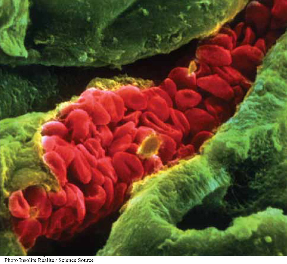
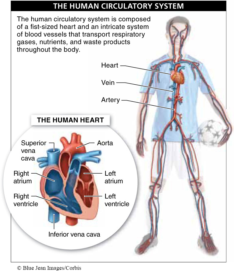
Clench your fist. That is the size of your heart. Now clench your fist and relax it a hundred thousand times. That is what your heart must do, every day, as it beats for 70 or more years. The human heart is at the center of our circulatory system, and it’s one of the most durable and reliable pumps ever produced (FIGURE 21-8).
In our earlier overview of the circulatory system, we saw that the four-
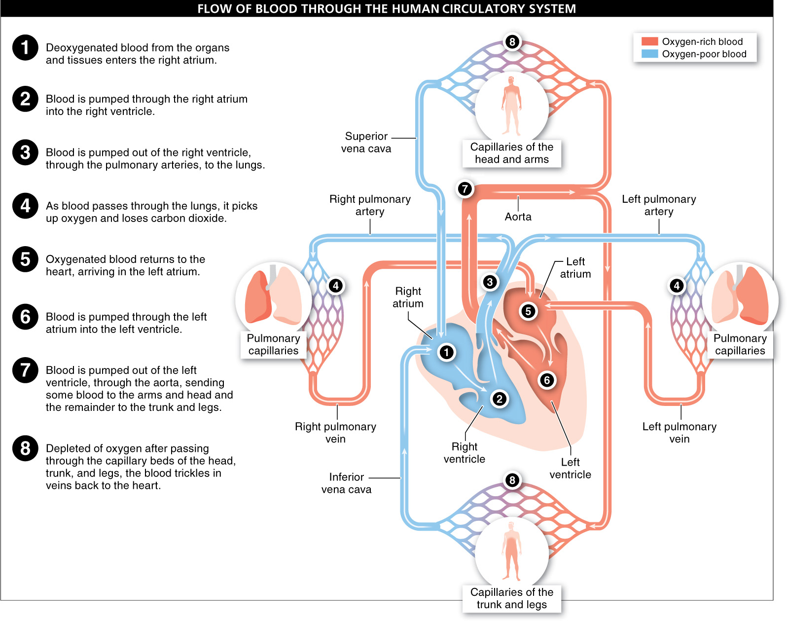
- 1. Deoxygenated blood from the organs and tissues enters the right atrium. The blood arriving from the lower half of the body enters through a large vein called the inferior vena cava. Blood arriving from the head and arms enters through the superior vena cava.
- 2. Most of the blood passes directly through the right atrium into the right ventricle. A contraction pushes the remainder of the blood in the right atrium into the right ventricle.
- 3. The contraction continues, pumping the blood out of the right ventricle and into the pulmonary artery. This large artery immediately forks, sending half of the blood to the left lung and half to the right lung.
- 4. As the blood passes through the pulmonary capillaries in the lungs, it picks up oxygen and loses carbon dioxide.
- 5. The oxygenated blood then enters the left and right pulmonary veins and returns to the left atrium of the heart.
- 6. Most of the blood passes directly through the left atrium and into the left ventricle. A contraction pushes the remaining blood from the left atrium into the left ventricle.
- 7. As the contraction continues, the oxygenated blood is pumped up and out of the ventricle at tremendous pressure into the aorta, the largest artery in the body. After making a sharp turn (called the aortic arch), the aorta forks and sends some blood to the arms and head and the remainder to the trunk and legs.
- 8. After passing through the capillary beds in the tissues of the head, arms, trunk, and legs, the blood moves at low pressure through the veins. This deoxygenated blood returns to the heart by way of the superior and inferior venae cavae and collects in the right atrium, and the cycle repeats.
836
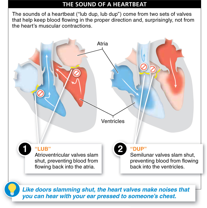
If you place your ear on the center of someone’s chest in a quiet room, you can hear the heart working: “lub dup, lub dup.” Over and over, the same two sounds. These are the sounds that a doctor hears when using a stethoscope. What is the source? It’s not the contraction of the heart. Surprisingly, the muscular contractions don’t make much noise. Rather, the sounds come from two sets of valves that help keep blood flowing in the proper direction (FIGURE 21-10).
The first set of valves is the atrioventricular (or AV) valves. Located between the atrium and ventricle on each side of the heart, these two valves allow blood to flow from the atrium to the ventricle. But when the ventricle contracts, these flaps of tissue slam shut, preventing blood from being pushed back into the atria: “lub.” With no other escape, the blood flows out through the pulmonary arteries on the right side and the aorta on the left. At these two primary exits from the heart are two more valves. These semilunar valves (so called because of their half-
What is the source of heart murmurs?
When the atrioventricular or semilunar valves do not completely close, some blood can squirt back through them, flowing in the wrong direction. The blood moving backward through the valve can be heard, with a stethoscope, making a buzzing or swishing noise. These noises are called heart murmurs. Most are not life-
TAKE-HOME MESSAGE 21.4
The human heart, at the center of our circulatory system, is an extremely durable pump. It sends blood on a figure 8, two-
When listening to a heartbeat, what are the “lub” and “dup” sounds that one hears?
The "lub" sound is caused by the atrioventricular valves slamming shut, preventing blood from flowing back into the atria. The "dup" sound is caused by the semilunar valves slamming shut, preventing blood from flowing back into the ventricles.