Vision: seeing is the perception of light by the brain.
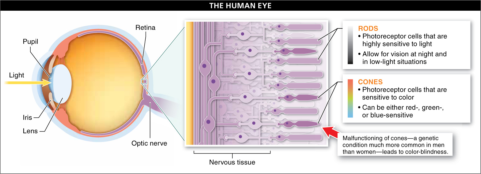
What we lack in smell proficiency, we more than make up for in visual acuity, a consequence of our image-forming, single-lens eyes. (This type of eye has evolved independently twice: in vertebrates and in a group of mollusks that includes the squid.) Light hits the eye and enters the eye’s interior through an iris. The pupil, an opening in the iris, opens and closes to control the amount of light that gets into the eye. Once the light is through the pupil, a lens focuses it onto the retina, nervous tissue containing light-sensitive cells called photoreceptor cells (FIGURE 23-22).
The retina lines the inner surface of the eye and transmits impulses to the vision center of the brain via the optic nerve. The two types of photosensitive cells in the retina are rods, which are highly sensitive to even tiny amounts of light and make it possible to see at night and in low-light situations, and cones, which are less sensitive to light and so are more effective during daylight. Humans have three kinds of cone cells: red-, green-, and blue-sensitive. Our ability to detect color is based on the combination of cones being stimulated. Some individuals (usually men) produce non-functioning red or green cones—the result of mutant genes. In either of these cases, the individual cannot distinguish red from green, so the person is said to be color-blind.
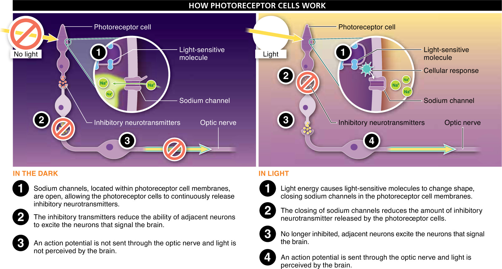
Vision is one of the senses in which our capabilities exceed those of most other animals. Still, as with nearly every trait, there are plenty of species—including many birds and some spiders—that have abilities that exceed ours. Despite the great diversity of light-sensing capabilities, the basic functioning of light-absorbing cells is quite consistent among all animals, although in some ways the process works in a fashion opposite to what we see for other types of sensory reception. Here’s how these cells work (FIGURE 23-23).
Figure 23-22The structures of the human eye.
Figure 23-23Photoreceptors responding to light enable vision.
Within the eye or other light-detecting structure, there are photoreceptor cells, with light-sensitive molecules embedded in their cell membranes. In the dark, a molecule binds to these photoreceptor cells, causing them to depolarize. They also continuously release a neurotransmitter that, like the depressing of a brake pedal, inhibits the ability of many adjacent neurons to send information to the brain. In a sense, darkness blocks the ability of optic nerves to send visual signals to the brain.
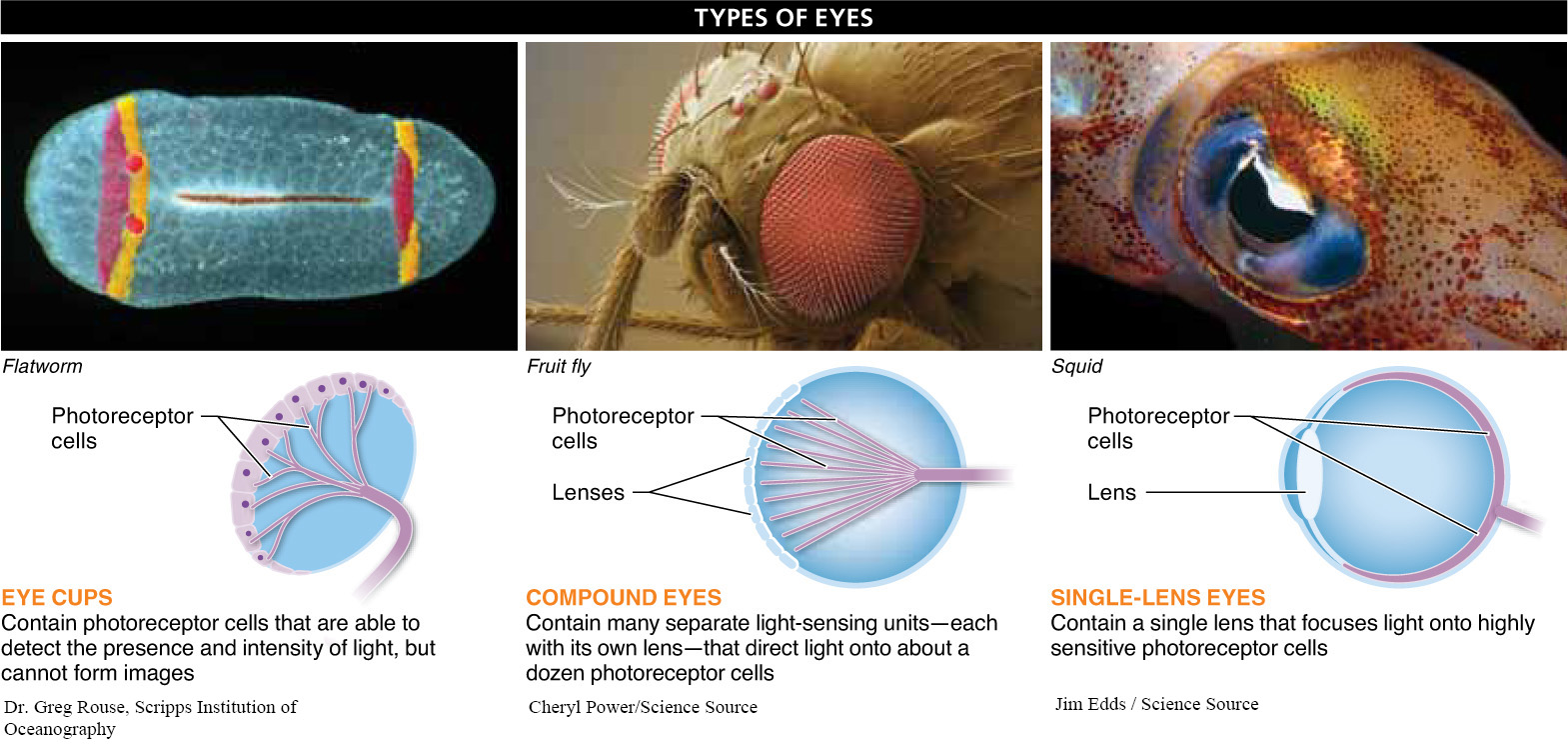
Figure 23-24Eye structure suits the needs of the organism.
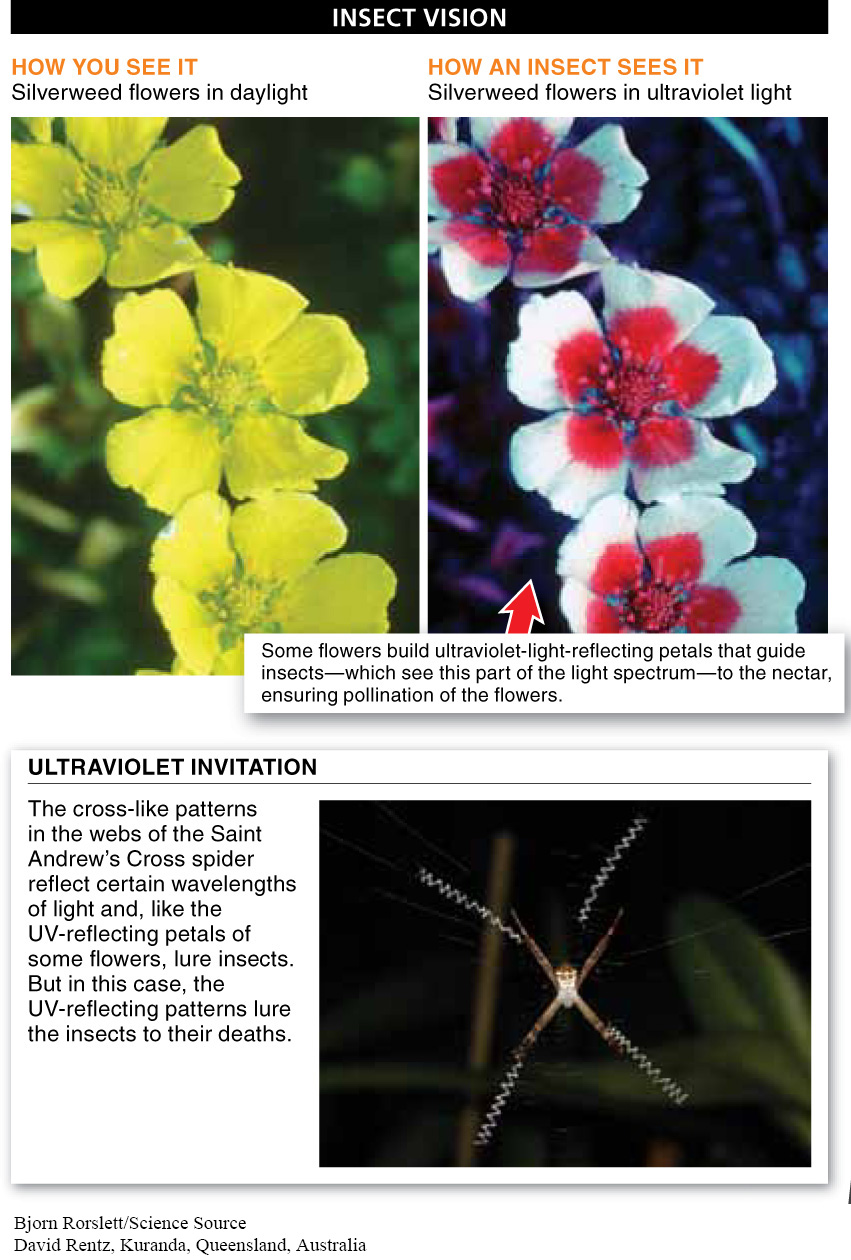
Figure 23-25Insects see things differently than we do.
When light hits one of the light-sensitive molecules in the membrane of a photoreceptor cell, the light energy causes some of the chemical bonds in the molecule to become stretched. (A similar capture of light energy occurs in plants during photosynthesis.) This closes sodium channels in the photoreceptor cell membranes and reduces the amount of inhibitory neurotransmitter that is released. Without the inhibitory neurotransmitter at the synapse, the adjacent neurons are able to excite the neurons that send visual signals to the brain. The particular wavelength perceived by the brain depends on the particular light-sensitive molecules stimulated.
In addition to the single-lens eye, two other types of eyes have evolved (FIGURE 23-24)—the eye cup and the compound eye. The simplest form is eye cups, found in the flatworms, such as planaria, which live in ponds and streams. Eye cups are made up of photoreceptors, and although they cannot form images, they are able to detect the presence and intensity of light. Unless the light is directly in front of the worm, it shines more intensely on one eye than the other.
937
938
This allows the animal to sense which direction the light is coming from. Usually, this information tells the animal which way to move: toward or away from the light. Planaria, for example, move away from the light.
Insects have much more refined visual capabilities (FIGURE 23-25). They possess compound eyes made of dozens to thousands of separate light-sensing units, each with its own image-forming lens that directs light onto about a dozen photoreceptors. The photoreceptors then send signals along neurons. When the signals reach the brain, the brain interprets these signals as an image. Because some insect photoreceptor cells—such as those in honeybees—contain a broader range of light-sensitive pigments than is found in humans, these insects can see wavelengths of light in the ultraviolet spectrum that are invisible to us.
Q
Insects don’t just fly into spiders’ webs accidentally. Sometimes they do it on purpose. Why?
Plants sometimes capitalize on this ability of insects to detect the ultraviolet spectrum by constructing flowers with UV-reflecting petals that look like the landing lights at an airport, guiding insects to their nectar reward (while ensuring pollination of the flowers). Devious spiders capitalize on this ability, too. Some build webs with UV-reflecting threads that trick insects into thinking the web is a flower. Once it flies into the web, the insect finds no nectar and ends up as a meal for the spider.
TAKE-HOME MESSAGE 23.11
Vision results from the stimulation of light-sensitive sensory neurons, called photoreceptor cells. The photoreceptor cells have a variety of molecules, embedded within their membranes, that are chemically altered by light. Signals are conveyed to the brain and interpreted as an image. The particular wavelength perceived depends on which version of the light-sensitive molecules in the photoreceptor cell membranes is stimulated.
Briefly describe the two types of photosensitive cells in the retina of the human eye.
Rods are highly sensitive to even tiny amounts of light. They make it possible to see at night and in low-light situations. Cones are less sensitive to light and more effective during daylight. Both have in common the use of sodium channels in their cell membranes and the use of inhibitory neurotransmitters to signal the brain; however, they use these processes differently.