
Running, flying, swimming, crawling, walking, digging, dancing. Animals, with very few exceptions, move. And in vertebrates, movement is generally initiated by cells of the nervous system that stimulate muscle contractions that pull on a rigid skeletal system. In this section, we examine the three types of muscle tissue, how contractions are generated within that tissue, and how input from the nervous system can initiate and control muscle contractions. We also investigate how the size and strength of muscles reflect their use and how physical training can increase muscle strength.
As we saw in Section 20-
- 1. Skeletal muscle is attached to bones by connective tissue and is controlled by individual neurons attached to each muscle fiber. It accounts for most of the movement we see in vertebrates and makes up about 40% of human body weight. The muscular system of humans contains about 700 skeletal muscles in all.
- 2. Cardiac muscle causes the heart to pump blood through the body. Not initiated by the nervous system or under conscious control, cardiac muscle contractions and relaxations occur continuously throughout life, with the muscle cells containing more energy-
releasing mitochondria than other types of muscle cells. - 3. Smooth muscle, which also is not under conscious control, surrounds blood vessels and many internal organs. In some cases, smooth muscle contractions are stimulated by the autonomic nervous systems, and in other cases, smooth muscle is able to contract without any stimulation from the nervous system. Compared with the other muscle types, smooth muscle generates slower contractions, which can gradually move blood, food, or other substances through vessels and organs.
944
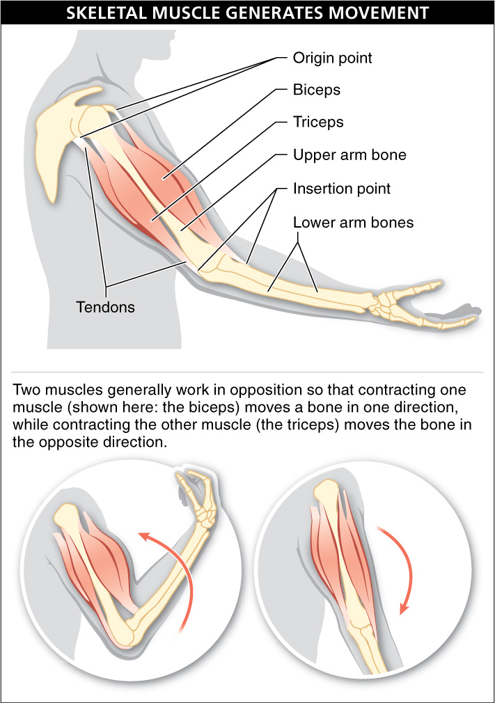
Our focus here is on skeletal muscle. To facilitate movement, skeletal muscles are attached to bones by connective tissue. Often, a muscle tapers at each end into tendons, which attach the muscle at the two ends—


945
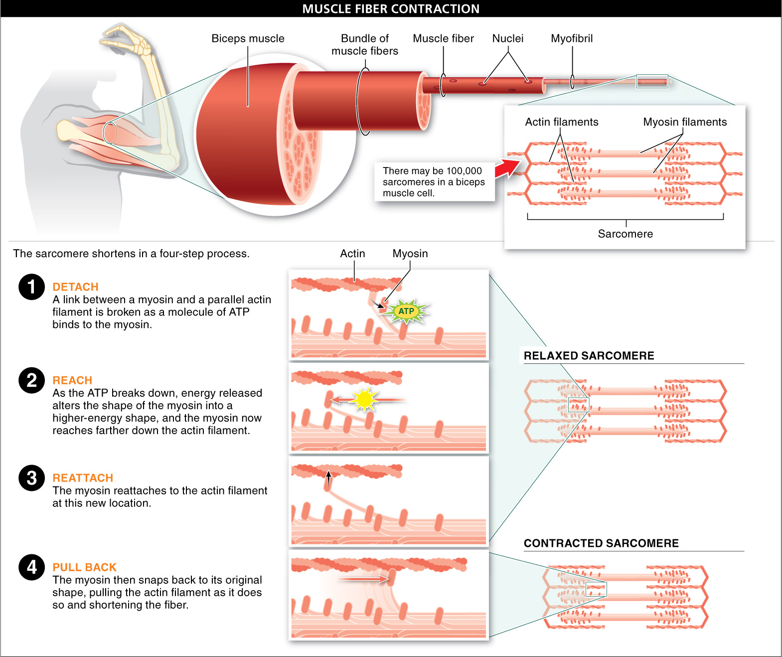
A skeletal muscle is made up of a bundle of fibers. Each fiber is a single cell, but has multiple nuclei and numerous myofibrils, cylindrical organelles that shorten when they contract. A myofibril contains repeating units called sarcomeres, which are where the contraction takes place.
The sarcomeres are composed of large numbers of long filaments, overlapping and parallel to each other. The filaments come in two types: thin filaments made mostly from the protein actin, and thick filaments made mostly from the protein myosin. There may be 100,000 sarcomeres in a biceps muscle cell.
The sarcomere shortens in a four-
- 1. Detach. A link between a myosin filament and an actin filament parallel to it is broken as a molecule of ATP binds to the myosin.
- 2. Reach. As the ATP breaks down, energy released alters the shape of the myosin into a higher-
energy shape, much like bending a twig— the bent twig is in a higher- energy shape because it can release energy as it snaps back to its original shape. - 3. Reattach. In its altered shape, the myosin reaches farther down the actin filament, where it reattaches.
- 4. Pull back. The myosin then snaps back to its original shape, pulling the actin filament as it does so and thus shortening the fiber. This last step is considered the “power stroke.”
As the contraction process occurs across numerous sarcomeres in a muscle cell, the entire muscle can shorten. From the perspective of energy, the potential energy of ATP is converted to the kinetic energy of sarcomere shortening, which can do work (for instance, as your arm curls a dumbbell).
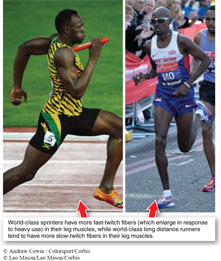
Marathon runners tend to have smaller leg muscles than sprinters. Why?
When a muscle contracts, the duration between a contraction and a relaxation is called a twitch. Muscle fibers within a muscle vary in how quickly they can twitch, with two general types. Fast-
When muscles are used for high-
TAKE-HOME MESSAGE 23.15
Muscle tissue—
Describe the four-
First, a link between an actin fiber and a parallel myosin fiber is broken as an ATP molecule binds to the myosin. Then, as the ATP molecule is broken down, energy released alters the shape of the myosin. The myosin in its altered shape is then reattached to the actin, though at a different location. Finally, the myosin snaps back to its original shape, pulling the actin fiber in the process and shortening the fiber.
946