
We owe a great deal to giant squids. Much of what we know about human nervous systems has come from studying squid neurons. Neurons function almost exactly the same in all animals, and giant squids just happen to also have giant neurons. While a human neuron might be 0.02 millimeter in diameter, a squid’s may be more than 100 times wider, up to double the thickness of a human hair. This makes squid neurons much easier to manipulate and observe. Regardless of the species under observation, each neuron has one axon. This projection leaves the cell body and can extend several feet or more. At its end, the axon branches into several (or hundreds of) axon terminals, also called terminal buttons, which are knob-
926
We’ve said that axons are like the electrical wires of the nervous system. In your home, electrical wires are usually covered with rubber. Besides reducing the likelihood of someone receiving an electric shock from the current running through the wire, this insulation also has another function: it causes the electrical charge to move through the wire more quickly and reduces dissipation of the signal. Axons are similarly insulated, by a fatty coating called the myelin sheath, preventing the action potential from weakening as it travels down the axon. But, unlike the solid rubber coating of an electrical wire, a myelin sheath has gaps that assist in propagating an action potential. Ion channels are concentrated at the gaps in the myelin sheath and allow sodium ions to rush into the axon. The rushing ions make the cell’s charge sufficiently positive in that region to cause the opening of ion channels at the next gap in the myelin sheath, thereby propagating the action potential along the axon.
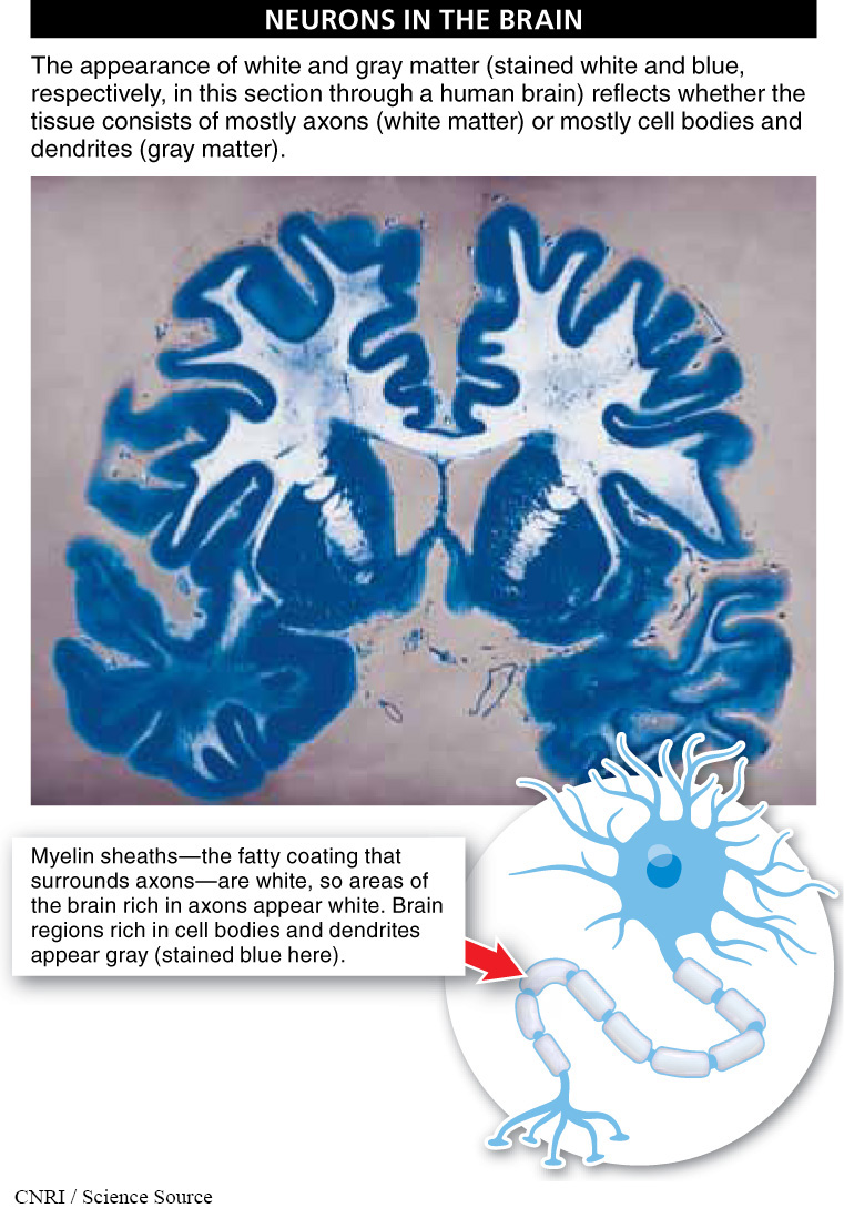
Because the fatty myelin is white, in cross sections of the brain, those areas where axons are densely packed together appear white (FIGURE 23-10). Regions of the brain with many cell bodies and dendrites appear gray. These different regions are often referred to as white matter and gray matter.
What do you think would happen to signals traveling down an axon in your leg if the myelin sheath insulator were not there? You can observe this effect by watching babies when they first start trying to walk, at around 10 or 11 months of age. At that time, myelin hasn’t completely formed around all of their axons, and consequently, their gross motor control isn’t very good (FIGURE 23-11). So, although the baby’s brain may be saying “Walk!” the signal never makes it to the leg muscles. Walking is especially difficult to master because the areas of the brain that control the feet and legs don’t develop until after the areas controlling the rest of the body.

Some symptoms of multiple sclerosis resemble difficulties that babies have when learning to walk. Why?
Multiple sclerosis (MS) is a disease that affects the central nervous system. In MS, myelin is gradually lost, leaving scar tissue called sclerosis. As myelin is lost, the neuron gradually loses its ability to conduct electrical impulses. This makes it progressively more difficult for the brain to send signals to muscles—
927
TAKE-HOME MESSAGE 23.5
Each neuron has one axon, a projection that extends from the cell body, sometimes several feet or more. At its end, the axon branches into numerous axon terminals (terminal buttons), positioned close to a muscle cell or a gland or to the dendrites of another neuron. In response to an action potential, the axon terminals release chemicals into the extracellular space, potentially influencing adjacent cells.
How is the myelin sheath of an axon like the rubber coating of an electrical cord?
Axons are like the electrical wires of the nervous system, propagating an electrical charge (the action potential) down their length. Continuing with the analogy, electrical wires are usually covered with rubber to allow the electrical charge to move more quickly through the wire and also prevent dissipation of the charge. Similarly, axons are insulated by a fatty coating called the myelin sheath, which prevents the action potential from weakening as it travels down the axon.
928