An action potential moving rapidly down an axon quickly runs out of axon. This is the end of one neuron, but not necessarily the end of the signal. As we’ve seen, the end of an axon—

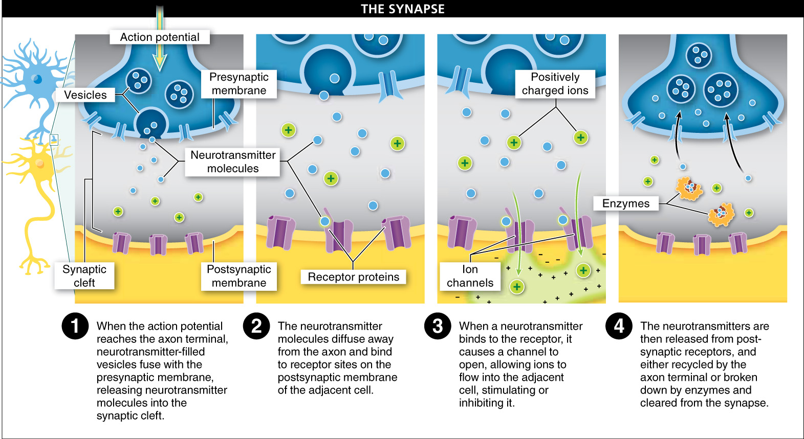
- 1. Sacs called vesicles release neurotransmitters into the synaptic cleft. When the action potential reaches the axon terminal, it causes little sacs called vesicles to merge with the axon’s cell membrane, called the presynaptic membrane. The sacs open up and release their contents, chemical messengers called neurotransmitters, into the synaptic cleft, the space between the axon and the cell receiving the signal (muscle cell, gland, or neuron).
- 2. Neurotransmitter diffuses and binds to nearby receptor sites. As the neurotransmitter molecules float around in the fluid in the synaptic cleft, they diffuse away from the axon until they bump into the adjacent neuron, muscle cell, or gland. Some of the neurotransmitter molecules attach to receptor sites on the postsynaptic membrane of the adjacent cell (called the postsynaptic cell).
- 3. Gates open in the postsynaptic cell membrane, and the signal enters the postsynaptic cell. When neurotransmitter binds to a receptor in the postsynaptic cell membrane, this causes a gate to open, which allows ions (often sodium ions) to flow into the cell. The resulting chemical change in the postsynaptic cell can cause an electrical change and, consequently, initiate an action potential (if the postsynaptic cell is a neuron), a contraction (if it’s a muscle cell), or a secretion (if it’s a gland).
- 4. Neurotransmitter is released from the postsynaptic cell receptors and recycled or broken down. The receptors then release the bound neurotransmitter molecules back into the fluid in the synaptic cleft. Eventually, the neurotransmitter molecules return to the presynaptic membrane and are recycled, or they are broken down within the synaptic cleft by enzymes, clearing out the area so that the synapse can be used again.
929

A contraction may occur if the postsynaptic cell is a muscle cell, and a secretion may occur if the postsynaptic cell is a gland, but how do neurotransmitters affect a neuron? In some cases, the released neurotransmitters are excitatory and excite the next neuron, increasing the likelihood that it will fire its own action potential. In other cases, the neurotransmitters are inhibitory and reduce the likelihood that the next cell will produce an action potential. And for some neurotransmitters, whether it is excitatory or inhibitory depends on the receptor. With the giant web of connections between neurons—
It might seem odd that the total of all the signals might direct the cell not to fire. However, the capacity to stop the signal enables the nervous system to modulate and filter some of the overwhelming amount of sensory information coming into the brain. It’s like call-
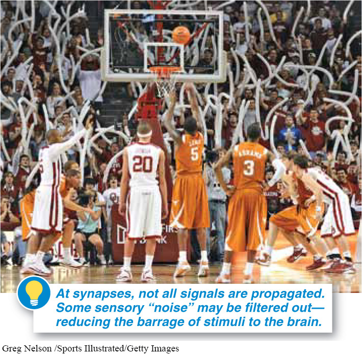
This “filtering” characteristic of the nervous system is one of the main reasons that organisms don’t have single, long neurons running from their sensory receptors right to their brain or muscles. It’s not always best to have every signal propagated (FIGURE 23-13).With continuous stimulation, too, most neurons gradually reduce the amount of neurotransmitter they release, and thus reduce the strength of the signal. It’s as if the neurons are saying, “Enough already. We get the message.”
TAKE-HOME MESSAGE 23.6
At the synapse, a neuron interacts with other cells. In response to an action potential, neurotransmitters are released into the synaptic cleft, diffuse, and may bind to receptors on an adjacent neuron, muscle cell, or gland, potentially stimulating an action potential, muscle contraction, or secretion. Neurotransmitters may then be taken back in by the axon terminal or enzymatically broken down in the synaptic cleft.
How does a neuron transmit its signal to another cell?
The action potential travels throughout the length of the axon and reaches the axon terminal, a site near another cell. The action potential causes sacs filled with neurotransmitters to fuse with the cell membrane, releasing these neurotransmitters from the neuron into the synaptic cleft, the space between the neuron and another cell. These neurotransmitters may bind to receptors in this adjacent (postsynaptic) cell, causing certain ion channels to open. Ions flow into this adjacent cell, initiating a response. The receptors then release the bound neurotransmitter, which is recycled or broken down.