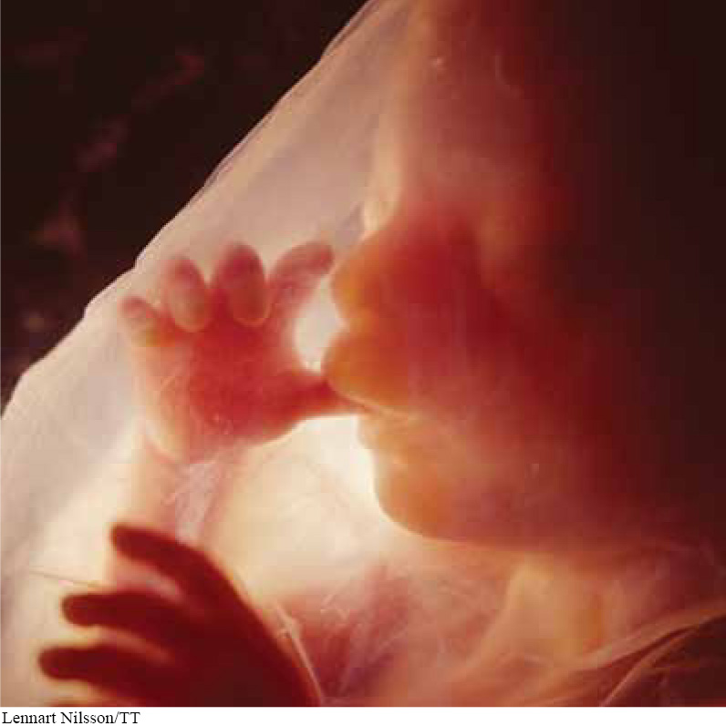
How do you build a complete human, with specialized cells, tissues, and organs, when at fertilization there is just a zygote, a single cell that doesn’t resemble a human at all? The field of developmental biology seeks to understand and explain how a complex organism develops from a single cell. To be sure, the genes in that initial cell are important, and the environmental conditions in which the cell develops are also critical. These factors interact as the development of humans and most other vertebrates proceeds in a carefully coordinated sequence of three stages: cleavage, gastrulation, and neurulation.
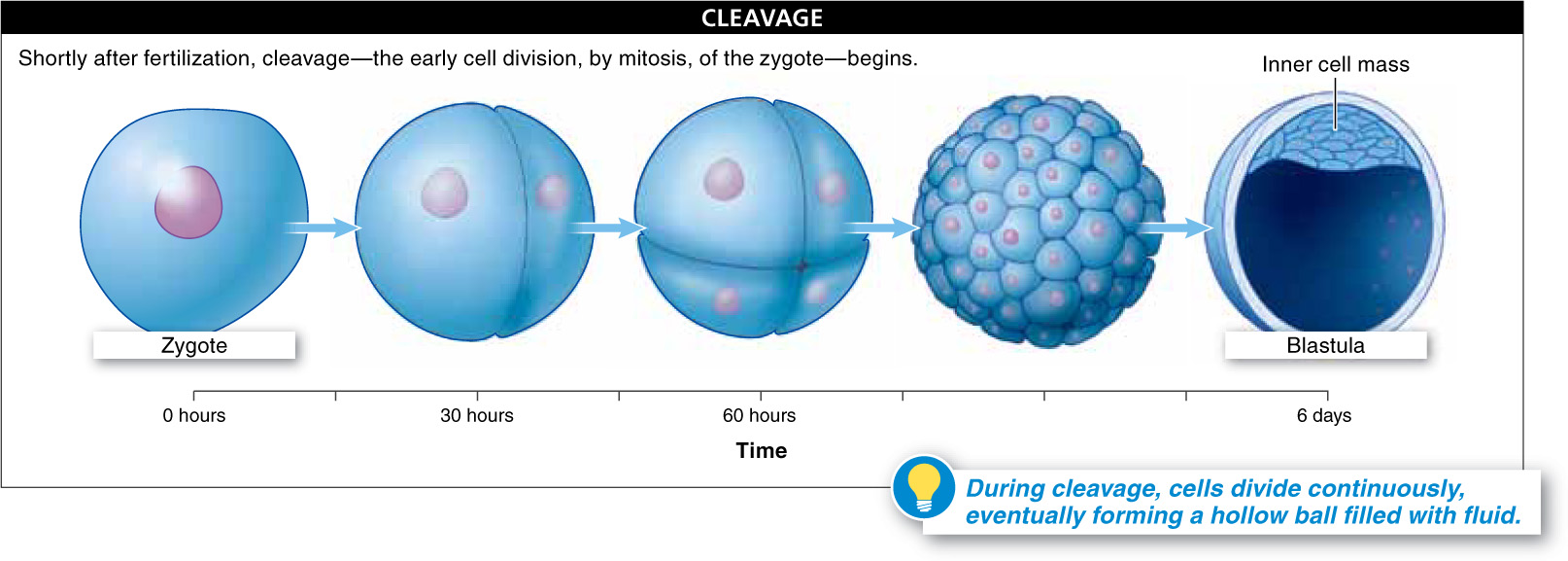
Cleavage The first phase, cleavage, is the early cell division, by mitosis, of the zygote, beginning shortly after fertilization (FIGURE 25-23). It starts about 30 hours after fertilization, as the zygote divides into two cells. Then, after another 30 hours or so, it divides again, becoming four cells, and then again, through numerous divisions. Although the number of cells increases significantly during cleavage, there is little or no growth in overall size. Rather, the fertilized egg is partitioned into more and more cells that are smaller and smaller. These multiple cell divisions are fueled by nutrients that were in the egg. At the cleavage stage, it is difficult to distinguish between species as dissimilar as sea urchins and humans.
1028
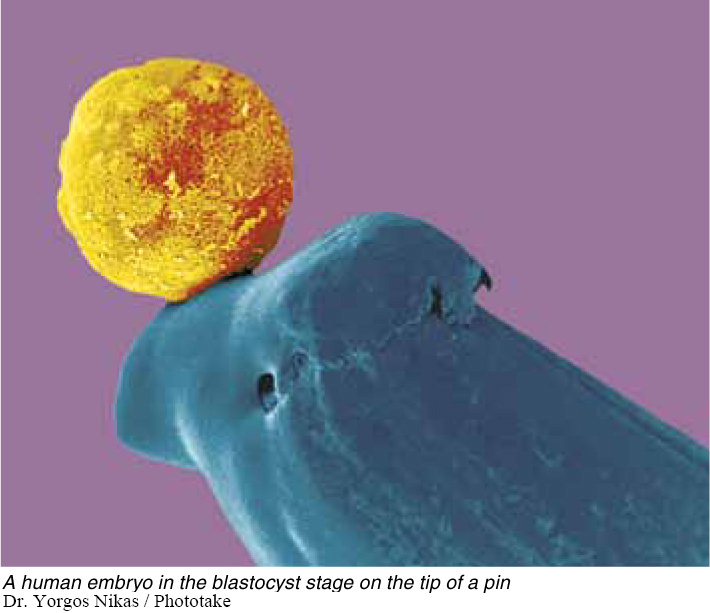
After about six days of continuous cell division, the cells form a hollow ball, called a blastula—the mammalian version is called a blastocyst—of approximately a thousand cells. The cells secrete fluid into the center of the ball, where there is an inner mass of cells that form the embryo. The outer cells also produce human chorionic gonadotropin (hCG), which keeps the corpus luteum from disintegrating. And the continued maintenance of the corpus luteum keeps progesterone levels high.
The blastocyst grows rapidly and forms membranes that surround and protect it. These include the amnion, which surrounds the embryo, and the chorion, which, along with the endometrium, forms the placenta, the structure that connects the developing embryo to the wall of the uterus (see Figure 25-29). The placenta is packed with blood vessels that bring nourishment to the embryo and remove wastes generated by the embryo (FIGURE 25-24).
Gastrulation In the second phase of development, gastrulation, three distinct germ layers of tissue form. Picture a beach ball—
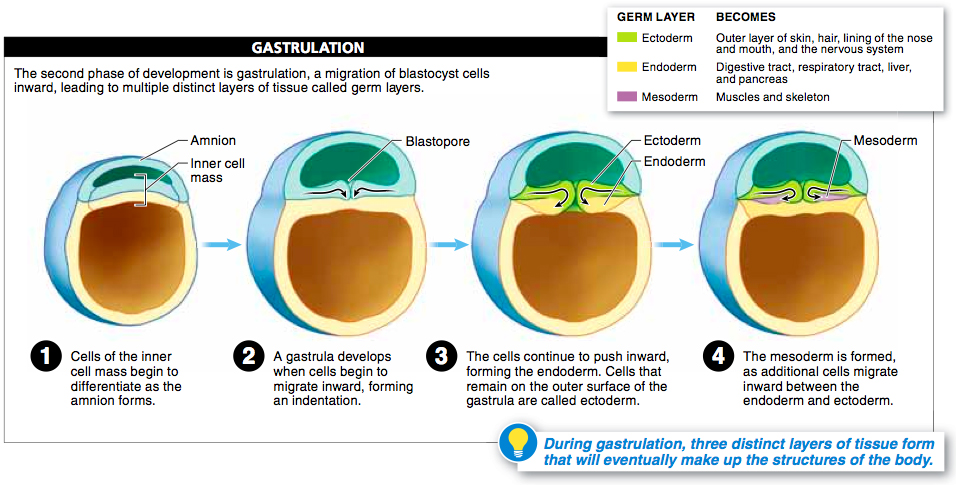
Three distinct layers of tissue form during gastrulation. The cells in these layers have not yet differentiated (that is, they all look similar to one another), but they have become “determined,” which means that the type of tissue they will become, their ultimate developmental fate, is irreversibly decided. Prior to determination, cells are referred to as “totipotent” and have the capacity to develop into any type of tissue in the body.
Here’s a summary of what happens during gastrulation.
- The blastocyst begins to fold inward, forming an indentation, the blastopore.
- As the cells continue to push inward, they are called endoderm, and they form the lining of what will eventually become the digestive tract. Endoderm cells will also form the liver, the pancreas, and the lining of the respiratory tract. At this point, the entire mass of cells is called a gastrula.
- The cells that remain on the outer surface of the gastrula are called ectoderm and will ultimately form the outer layer of skin, the hair, the lining of the nose and mouth, and the nervous system.
- Some additional cells migrate inward, between the endoderm and ectoderm, forming a third type of tissue, called mesoderm, which gives rise to the muscles and skeleton.
The specific details of gastrulation differ slightly from one species to the next. The general outcome, however—
Neurulation In the third week of embryonic development, the three types of tissue formed during gastrulation begin to develop into the various organs and tissues. At this time, within the mesoderm and running the length of the embryo, just above the digestive tract, the notochord forms. The notochord is a flexible rod that develops in all chordates and gives support to surrounding tissue. In vertebrates, the notochord’s function is ultimately taken over by the backbone.
Just above the notochord, ectoderm folds in for the entire length of the embryo, first forming a groove and then becoming a long hollow tube, called the neural tube. The process is referred to as neurulation, and the resulting neural tube ultimately develops into the entire nervous system, including the brain and the spinal cord (FIGURE 25-26).

1029
As the neural tube is developing, blocks of mesoderm tissue, called somites, begin to form down the length of the embryo. These somites will form the vertebrae and the muscles. Next to the somites, spaces form that will become the body cavity, or coelom (pronounced-
1030
Other cells may be induced to develop into one particular cell type by neighboring cells that send signals, possibly proteins, to turn certain genes on or off. This process was demonstrated dramatically by researchers who transplanted cells from the top of a newt blastopore into a different developing embryo. In the recipient embryo, the blastopore cells induced the development of a second neural tube!
TAKE-HOME MESSAGE 25.12
Soon after fertilization, cleavage takes place, and many rapid cell divisions occur without overall growth. Then, in gastrulation, a gut begins to form, along with three distinct germ layers with specific developmental fates. In neurulation, mesoderm forms a supporting rod called the notochord, and above that, an infolding of ectoderm forms a neural tube, which will become the brain and spinal cord.
What structures are formed from the endoderm?
The innermost layers of cells form the endoderm, which become the digestive tract, liver, pancreas, and the lining of the respiratory tract.