Mitosis is an all-
Meiosis, then, starts with a diploid cell—

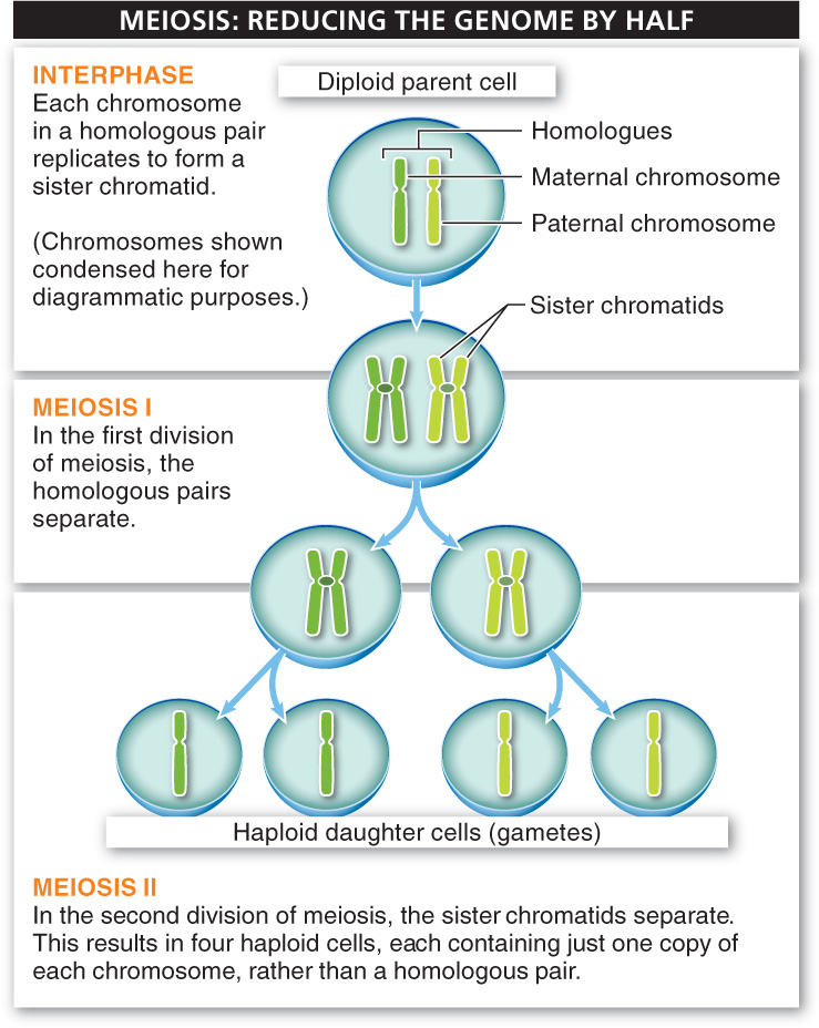
Before meiosis can occur (and just as we saw with mitosis), each of these 46 chromosomes is duplicated during the cell’s interphase. This means that when meiosis begins, we have 92 (2 × 46) strands of DNA, after replication.

Unlike mitosis, which has only one cell division, cells undergoing meiosis divide twice. In the first division, the homologues separate. In other words, for each of the 23 chromosome pairs, the maternal sister chromatid pairs and the paternal sister chromatid pairs separate into two new cells. In the second division, each of the two new cells divides again, so that each of the four daughter cells contains a single chromosome from the homologous pair. At the end of meiosis, there are four new cells, each of which has 23 strands of DNA—
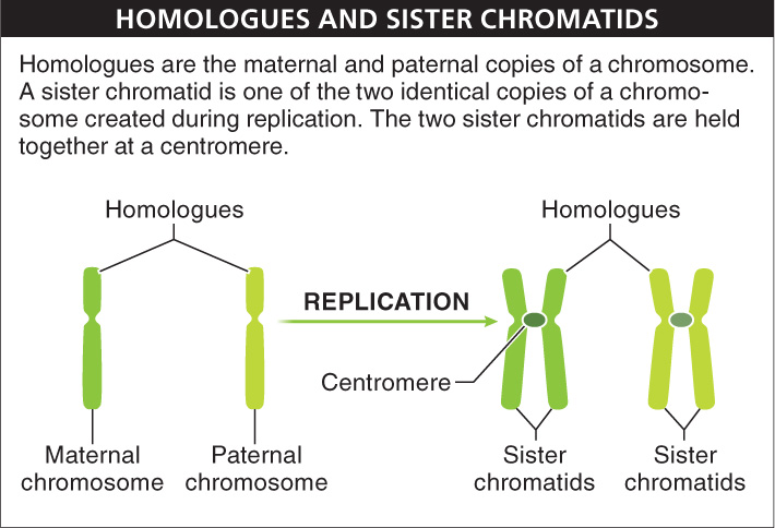
Interphase: In Preparation for Meiosis, the Chromosomes Replicate Before meiosis begins, preparations for cell division occur that are similar to those taking place in the interphase of mitosis in the somatic cell cycle. In particular, every chromosome creates an exact duplicate of itself by replication. The chromosomes that were each a single, long, linear piece of genetic material become a pair of identical long, linear pieces, held together at the centromere.
23
Meiosis begins following this replication step during interphase; it proceeds through eight steps (FIGURE 6-24).

Meiosis Division I: The Homologues Separate The first meiotic division takes place in four stages.
1. Prophase I: chromosomes condense and crossing over occurs. This is by far the most complex of all the phases of meiosis. As in mitosis, it begins with all of the replicated genetic material condensing.
As the sister chromatids become shorter and thicker, the homologous chromosomes come together. This is where the process diverges from mitosis. The homologous chromosomes (each of which has become a pair of identical chromatids) line up, touching. Under a microscope, the two homologous pairs of sister chromatids appear as pairs of X’s lying on top of each other.
At this point, the sister chromatids that are next to each other do something that makes every sperm or egg cell genetically unique: they swap little segments of DNA. In other words, when this is happening inside you, some of the genes that you inherited from your mother get swapped onto the strand of DNA you inherited from your father, and the corresponding bits from your father are inserted into the DNA strand from your mother. This event is called genetic recombination (or, more often, just recombination) or crossing over, and it can take place at several spots (up to dozens) on each chromatid. As a result of crossing over, every sister chromatid ends up having a unique mixture of your genetic material. Note that crossing over occurs only during the production of gametes, in meiosis. It does not occur during mitosis. Following crossing over, the nuclear membrane disintegrates.
We explore crossing over in more detail in Section 6-
2. Metaphase I: all chromosomes line up along the center of the cell. After crossing over, each pair of homologous chromosomes (that is, the pairs of X’s lying on top of each other) moves to the center of the cell, pulled by the spindle fibers to form the arrangement called the metaphase plate. Remember that each pair of homologous chromosomes includes the maternal and paternal versions of the chromosome (with crossed-
3. Anaphase I: homologues are pulled to either side of the cell. This phase is the beginning of the first cell division that occurs during meiosis. In anaphase, the homologues are pulled apart toward opposite poles of the cell. One of the homologues (consisting of two sister chromatids) goes to one pole, the other to the opposite pole. At this point, something else occurs that, along with crossing over, contributes to making all the products of meiosis genetically unique. The maternal and paternal sister chromatid pairs are pulled to the ends of the cell in a random fashion called random assortment. As a result of random assortment, the pairs of sister chromatids gathered at each pole are a mix of maternal and paternal sister chromatids.
Imagine all the different combinations that can occur in a species with 23 pairs of chromosomes. For chromosome pair 1, perhaps the maternal homologue goes to the “top” pole and the paternal to the “bottom” pole. And for pair 2, perhaps the maternal homologue also goes to the top and the paternal to the bottom. But maybe for pair 3, it is the paternal homologue that goes to the top and the maternal homologue to the bottom. For each of the 23 pairs, you cannot predict which will go to the top and which will go to the bottom. There is a huge number of possible combinations. (Can you figure out how many? Here’s a hint: if there were 2 chromosome pairs, the answer would be 4; if there were 3 chromosome pairs, the answer would be 8.)
4. Telophase I and cytokinesis: nuclear membranes reassemble around sets of sister chromatid pairs, and two daughter cells form. After the pairs of chromatids arrive at the two poles of the cell, nuclear membranes re-
Meiosis Division II: Separating the Sister Chromatids There is a brief interphase after the first division of meiosis. In some organisms, the DNA molecules (now in the form of chromatid pairs) briefly uncoil and fade from view. In others, the second part of meiosis begins immediately. It is important to note that in the brief interphase before prophase II, there is no replication of any of the chromosomes. The second part of meiosis, like the first division of meiosis, is a four-
24
5. Prophase II: chromosomes re-
6. Metaphase II: sister chromatid pairs line up at the center of the cell. In each of the two daughter cells, the sister chromatid pairs (each pair appearing as an X) move to the center of the cell, pulled by spindle fibers attached to the centromere, where the sister chromatids are held together. The congregation of all the genetic material in the center of each daughter cell is visible as a flat metaphase plate.
7. Anaphase II: sister chromatids are pulled to opposite sides of the cell. This phase starts with 46 pieces of DNA in each cell created by meiosis I: that is, 23 sister chromatid pairs. During this anaphase, the fibers attached to the centromere begin pulling each chromatid in the 23 sister chromatid pairs toward opposite poles of the daughter cell. When anaphase II is finished, each of what will become the four daughter cells (you could think of them as granddaughter cells!) has one single copy of each of the 23 chromosomes.
8. Telophase II and cytokinesis: nuclear membranes reassemble and the two daughter cells pinch into four haploid gametes. After the sister chromatids for all 23 chromosomes have been pulled to opposite poles, the cytoplasm divides. The cell membrane pinches each cell into two new daughter cells, nuclear membranes begin to re-
In humans, the outcome of one diploid cell undergoing meiosis is the creation of four haploid daughter cells, each with just one set of 23 individual chromosomes. These chromosomes contain a combination of traits from the individual’s diploid set of chromosomes.
TAKE-HOME MESSAGE 6.10
Meiosis in animals occurs only in gamete-
Compare the events of metaphase I and anaphase I of meiosis to those of metaphase II and anaphase II of meiosis.
During metaphase I, homologous pairs of chromosomes move toward the center of the cell (referred to as the metaphase plate) and line up. In anaphase I, the homologues separate and are pulled to opposite poles of the cell. The sister chromatid pairs are pulled to the ends of the cell at random, resulting in a mix of maternal and paternal genetic material. In metaphase II, sister chromatid pairs line up at the center of the cell. Then, sister chromatids are pulled apart by spindle fibers toward opposite poles of the cell in anaphase II.
25