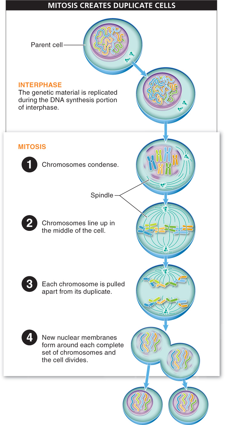
For mitosis to begin, the parent cell replicates its DNA, creating a duplicate copy of each chromosome. Once this task is completed, mitosis can take place. Mitosis occurs in just four steps, in which the now-
Before we explore the process of mitosis in more detail, it is helpful to clarify some of the terminology used to describe the important structures and processes.
14
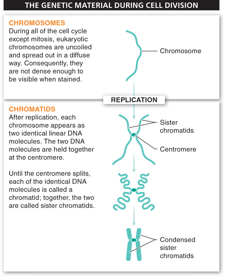
Generally, when you look at a cell through a microscope, you won’t see any chromosomes because the cells you’re looking at are in some part of their interphase. During all of the cell cycle except mitosis, chromosomes are uncoiled and spread out in a diffuse way, like a mass of spaghetti. And because they are so stretched out, they are not dense enough to be visible.
Before mitosis begins, two important events occur.
- 1. The chromosomes replicate, becoming two identical linear DNA molecules. The two DNA molecules are held together at a region called the centromere. Throughout mitosis, until the centromere splits, each of the identical DNA molecules is called a chromatid; together, the two are called sister chromatids (FIGURE 6-14).
- 2. The sister chromatids begin the process of condensation, in which they coil tightly and become compact—
in contrast to the uncondensed and tangled state of the chromosomes prior to replication, during most of interphase.
Animal chromosomes are linear. Why do they look like the letter X in pictures?
When the sister chromatids condense, they look like the letter X. This appearance can be confusing. As we saw earlier, chromosomes are not X-
Because a lot of room is required for the sister chromatids to separate, the membrane around the nucleus is dismantled and disappears early in mitosis. At the same time, a structure called the spindle, composed of proteins—
15
TAKE-HOME MESSAGE 6.6
Mitosis is the process by which cells duplicate themselves. Mitosis follows chromosome replication and leads to the production of two daughter cells from one parent cell.
Briefly describe what happens during the four steps of mitosis.
First, chromosomes condense. Then, chromosomes line up in the middle of the cell. Next, duplicate versions of chromosomes separate. Finally, each set of chromosomes is surrounded by a new nuclear membrane.