22.4 Programmed Cell Death Is an Integral Part of Development
An important aspect of development is the death of cells. Cell death shapes many body parts in the course of development: it is responsible for the disappearance of a tadpole’s tail during metamorphosis and causes the removal of tissue between the digits to produce the human hand. Cell death is also used to eliminate dangerous cells that have escaped normal controls (see the section on mutations in cell-cycle control and cancer in Chapter 23).
Apoptosis
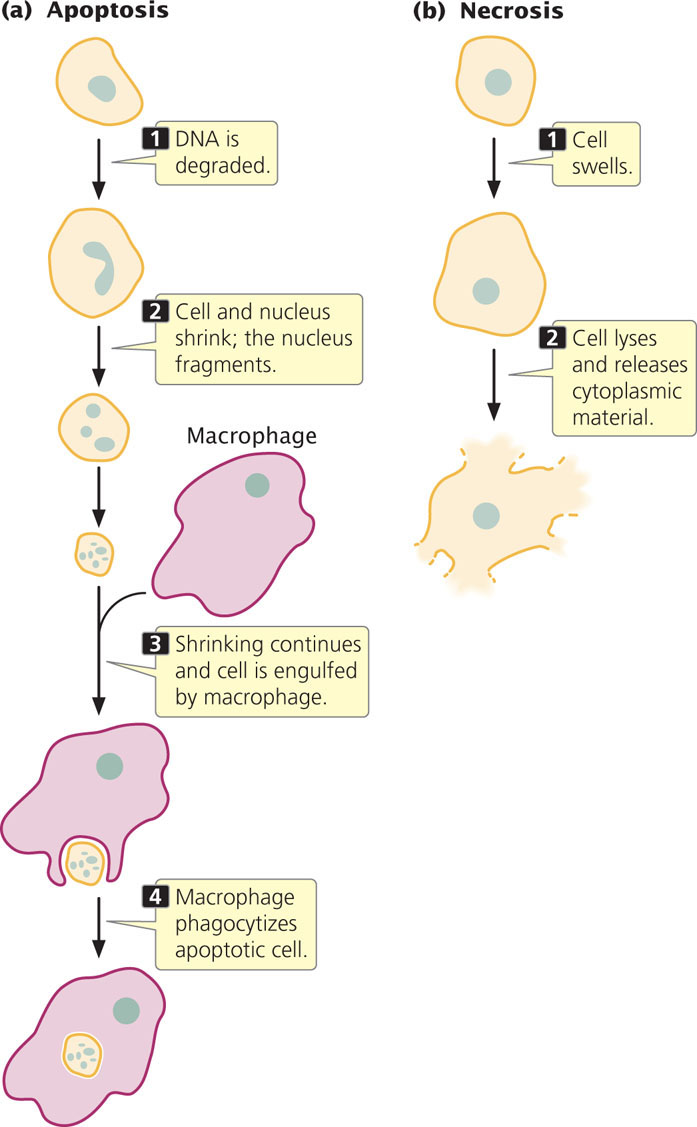
Cell death in animals is often initiated by the cell itself in apoptosis, or cellular suicide. In this process, a cell’s DNA is degraded, its nucleus and cytoplasm shrink, and the cell undergoes phagocytosis by other cells without any leakage of its contents (Figure 22.17a). Cells that are injured, on the other hand, die in a relatively uncontrolled manner called necrosis. In this process, a cell swells and bursts, spilling its contents over neighboring cells and eliciting an inflammatory response (Figure 22.17b). Apoptosis is essential to embryogenesis; most multicellular animals cannot complete development if the process is inhibited.
Regulation of Apoptosis
Surprisingly, most cells are programmed to undergo apoptosis and will survive only if the internal death program is continually held in check. Apoptosis is highly regulated and depends on numerous signals inside and outside the cell. Geneticists have identified a number of genes that have roles in various stages of the regulation of apoptosis. Some of these genes encode enzymes called caspases, which cleave other proteins at specific sites (after aspartic acid). Each caspase is synthesized as a large, inactive precursor (a procaspase) that is activated by cleavage, often by another caspase. When one caspase is activated, it cleaves other procaspases that trigger even more caspase activity. The resulting cascade of caspase activity eventually cleaves proteins essential to cell function, such as those supporting the nuclear membrane and cytoskeleton. Caspases also cleave a protein that normally keeps an enzyme that degrades DNA (DNase) in an inactive form. Cleavage of this protein activates DNase and leads to the breakdown of cellular DNA, which eventually leads to cell death.
Procaspases and other proteins required for cell death are continuously produced by healthy cells, and so the potential for cell suicide is always present. A number of different signals can trigger apoptosis; for instance, infection by a virus can activate immune cells to secrete substances onto an infected cell, causing that cell to undergo apoptosis. This process is believed to be a defense mechanism designed to prevent the reproduction and spread of viruses. Similarly, DNA damage can induce apoptosis and thus prevent the replication of mutated sequences. Damage to mitochondria and the accumulation of a misfolded protein in the endoplasmic reticulum also stimulate programmed cell death.
Apoptosis in Development
Apoptosis plays a critical role in development. As animals develop, excess cells are often produced and then later culled by apoptosis to produce the proper number of cells required for an organ or a tissue. In some cases, whole structures are created that are later removed by apoptosis. For example, early mammalian embryos develop both male and female reproductive ducts, but the Wolffian ducts degenerate in females and the Mullerian ducts degenerate in males. Apoptosis also plays an important role in the development of immune function (see section 22.6), where lymphocytes that recognize the body’s own cells normally undergo apoptosis so they do not attack self tissues. The immune system can also induce apoptosis in cells that are infected with viruses.
During embryonic development in Drosophila, large numbers of cells die. Three genes in Drosophila activate caspases that are essential for apoptosis: reaper (rpr), grim, and head involution defective (hid). Embryos possessing a deletion that removes all three genes exhibit no apoptosis and die in the course of embryogenesis with an excess of cells. Numerous other genes also affect the process of apoptosis.
Apoptosis is also crucial in metamorphosis, the process by which larval structures are transformed into adult structures. For example, the large salivary glands of larval fruit flies regress during metamorphosis. The hormone ecdysone stimulates metamorphosis, including the onset of apoptosis. Ecdysone induces the expression of rpr and hid and inhibits the expression of other genes, which then leads to apoptosis of salivary gland cells.
Apoptosis in Disease
The symptoms of many diseases and disorders are caused by apoptosis or, in some cases, its absence. In neurodegenerative disorders such as Parkinson disease and Alzheimer disease, symptoms are caused by a loss of neurons through apoptosis. In heart attacks and stroke, some cells die through necrosis, but many others undergo apoptosis. Cancer is often stimulated by mutations in genes that regulate apoptosis, leading to a failure of apoptosis that would normally eliminate cancer cells (see Chapter 23).
CONCEPTS
Cells are capable of apoptosis (programmed cell death), a highly regulated process that depends on enzymes called caspases. Apoptosis plays an important role in animal development and is associated with a number of diseases.
 CONCEPT CHECK 6
How does cell death from apoptosis differ from cell death from necrosis?