Meiosis
The words mitosis and meiosis are sometimes confused. They sound a bit alike, and both refer to chromosome division and cytokinesis. But don’t be deceived. The outcomes of mitosis and meiosis are radically different, and several unique events that have important genetic consequences take place only in meiosis.
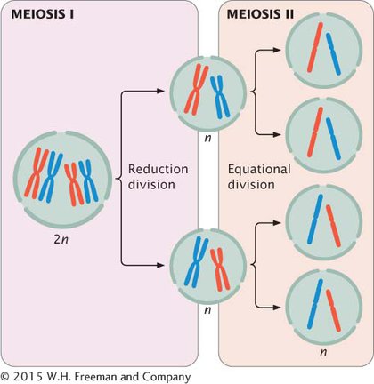
How does meiosis differ from mitosis? Mitosis consists of a single nuclear division and is usually accompanied by a single cell division. Meiosis, on the other hand, consists of two divisions. After mitosis, chromosome number in the newly formed cells is the same as that in the original cell, whereas meiosis causes chromosome number in the newly formed cells to be reduced by half. Finally, mitosis produces genetically identical cells, whereas meiosis produces genetically variable cells. Let’s see how these differences arise.
Like mitosis, meiosis is preceded by interphase, which includes G1, S, and G2 phases. Meiosis consists of two distinct processes: meiosis I and meiosis II, each of which includes a cell division. The first division, which comes at the end of meiosis I, is termed the reduction division because the number of chromosomes per cell is reduced by half (Figure 2.11). The second division, which comes at the end of meiosis II, is sometimes termed the equational division. The events of meiosis II are similar to those of mitosis. However, meiosis II differs from mitosis in that chromosome number has already been halved in meiosis I, and the cell does not begin with the same number of chromosomes as it does in mitosis (see Figure 2.11).
MEIOSIS I The stages of meiosis are outlined in Figure 2.12. During interphase, the chromosomes are relaxed and visible as diffuse chromatin. Prophase I is a lengthy stage in which the chromosomes form homologous pairs and crossing over takes place. First, the chromosomes condense, pair up, and begin synapsis, a very close pairing association. Each homologous pair of synapsed chromosomes, called a bivalent or tetrad, consists of four chromatids. The chromosomes become shorter and thicker. Crossing over, a process in which homologous chromosomes exchange genetic information, takes place. Crossing over generates genetic variation, as we will see shortly, and is essential for the proper alignment and separation of homologous chromosomes. Each location where two chromosomes cross is called a chiasma (plural, chiasmata). The centromeres of the paired chromosomes then move apart; the two homologs remain attached at each chiasma. Near the end of prophase I, the nuclear membrane breaks down and the spindle forms.
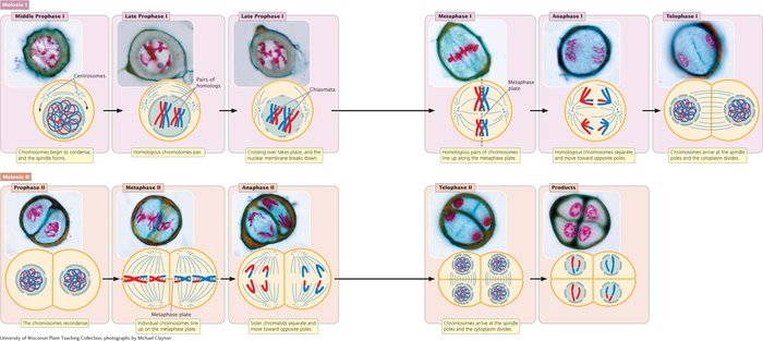
Metaphase I is initiated when homologous pairs of chromosomes align along the metaphase plate (see Figure 2.12). A microtubule from one pole attaches to one chromosome of a homologous pair, and a microtubule from the other pole attaches to the other member of the pair. Anaphase I is marked by the separation of homologous chromosomes. The two chromosomes of a homologous pair are pulled toward opposite poles. Although the homologous chromosomes separate, the sister chromatids remain attached and travel together. In telophase I, the chromosomes arrive at the spindle poles and the cytoplasm divides.
MEIOSIS II In the period between meiosis I and meiosis II, called interkinesis, the nuclear membrane re-
In anaphase II, the kinetochores of the sister chromatids separate and the chromatids are pulled to opposite poles. Each chromatid is now a distinct chromosome. In telophase II, the chromosomes arrive at the spindle poles, a nuclear envelope re-
| Stage | Major events |
|---|---|
| Meiosis I | |
| Prophase I | Chromosomes condense, homologous chromosomes synapse, crossing over takes place, nuclear envelope breaks down, and mitotic spindle forms. |
| Metaphase I | Homologous pairs of chromosomes line up on the metaphase plate. |
| Anaphase I | The two chromosomes (each with two chromatids) of each homologous pair separate and move toward opposite poles. |
| Telophase I | Chromosomes arrive at the spindle poles. |
| Cytokinesis | The cytoplasm divides to produce two cells, each having half the original number of chromosomes. |
| Interkinesis | In some types of cells, the spindle breaks down, chromosomes relax, and a nuclear envelope re- |
| Meiosis II | |
| Prophase II* | Chromosomes condense, the spindle forms, and the nuclear envelope disintegrates. |
| Metaphase II | Individual chromosomes line up on the metaphase plate. |
| Anaphase II | Sister chromatids separate and move as individual chromosomes toward the spindle poles. |
| Telophase II | Chromosomes arrive at the spindle poles; the spindle breaks down and a nuclear envelope re- |
| Cytokinesis | The cytoplasm divides. |
*Only in cells in which the spindle has broken down, chromosomes have relaxed, and the nuclear envelope has re-