In species with XX-XY sex determination, differences between males and females in their number of X chromosomes present a special problem in development. In females, there are two copies of the X chromosome and two copies of each autosome, so genes on the X chromosomes and on autosomes are “in balance.” In males, however, there is only a single X chromosome, while there are two copies of every autosome. Because the amount of a protein produced is often a function of the number of gene copies encoding that protein, males are likely to produce less of a protein encoded by X-linked genes than of a protein encoded by autosomal genes. This difference can be detrimental because protein concentration often plays a critical role in development.
Some animals have overcome this problem by evolving mechanisms to equalize the amount of protein produced by the single X and two autosomes in the heterogametic sex. These mechanisms are referred to as dosage compensation. In fruit flies, dosage compensation is achieved by a doubling of the activity of the genes on the X chromosome of males, but not of females. In placental mammals, the expression of dosage-sensitive genes on the X chromosomes of both males and females has been increased, and one of the X chromosomes is inactivated in females, so that the expression of X-linked and autosomal genes is balanced in both males and females.
For unknown reasons, the presence of sex chromosomes does not always produce problems of gene dosage, and dosage compensation of X-linked genes is not universal. A number of animals do not exhibit obvious mechanisms of dosage compensation, including butterflies and moths, birds, some fishes, and even the duck-billed platypus. As we will see in the next section, even in placental mammals a number of genes escape dosage compensation.
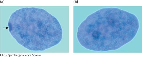
In 1949, Murray Barr observed condensed, darkly staining bodies in the nuclei of cells from female cats (Figure 4.11); these structures became known as Barr bodies. Mary Lyon proposed in 1961 that the Barr body was an inactive X chromosome. She suggested that within each female cell, one of the two X chromosomes is inactivated; which X chromosome is inactivated is random. Her hypothesis (now generally accepted for placental mammals) has become known as the Lyon hypothesis. If a cell contains more than two X chromosomes, all but one of them is inactivated. The number of Barr bodies present in human cells with different complements of sex chromosomes is shown in Table 4.3.
4.11 A Barr body is an inactivated X chromosome. (a) Female cell with a Barr body (indicated by arrow). (b) Male cell without a Barr body.
[Chris Bjornberg/Science Source.]
TABLE 4.3Number of Barr bodies in human cells with different complements of sex chromosomes
Sex chromosomes
Syndrome
Number of Barr bodies
XX
None
1
XY
None
0
XO
Turner
0
XXY
Klinefelter
1
XXYY
Klinefelter
1
XXXY
Klinefelter
2
XXXXY
Klinefelter
3
XXX
Triple-X
2
XXXX
Poly-X female
3
XXXXX
Poly-X female
4
Page 87
As a result of X inactivation, female placental mammals are functionally hemizygous at the cellular level for X-linked genes. In females that are heterozygous at an X-linked locus, approximately 50% of the cells express one allele and 50% express the other allele; thus, proteins encoded by both alleles are produced, although not within the same cell. This functional hemizygosity means that cells in females are not identical with respect to the expression of the genes on the X chromosome; females are mosaics for the expression of X-linked genes.
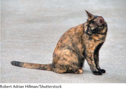
Random X inactivation takes place early in development—in humans, within the first few weeks of development. After an X chromosome has become inactive in a cell, it remains inactive in that cell and in all somatic cells that descend from that cell. Thus, neighboring cells tend to have the same X chromosome inactivated, producing a patchy (mosaic) pattern of expression of X-linked characteristics in heterozygous females.
This patchy distribution of gene expression can be seen in tortoiseshell (Figure 4.12) and calico cats. Although many genes contribute to coat color and pattern in domestic cats, a single X-linked locus determines the presence of orange color. There are two possible alleles at this locus: X+, which produces nonorange (usually black) fur, and Xo, which produces orange fur. Males are hemizygous and thus may be black (X+Y) or orange (XoY), but not black and orange. (Rare tortoiseshell males can arise from the presence of two X chromosomes, X+XoY.) Females may be black (X+X+), orange (XoXo), or tortoiseshell (X+Xo), with the tortoiseshell pattern arising from a patchy mixture of black and orange fur. Each orange patch is a clone of cells derived from an original cell in which the black allele was inactivated, and each black patch is a clone of cells derived from an original cell in which the orange allele was inactivated.
4.12 The patchy distribution of color on tortoiseshell cats results from the random inactivation of one X chromosome in females.
[Robert Adrian Hillman/Shutterstock.]
The Lyon hypothesis suggests that the presence of variable numbers of X chromosomes should not affect the phenotype in mammals because any X chromosomes in excess of one should be inactivated. However, persons with Turner syndrome (XO) differ from XX females, and those with Klinefelter syndrome (XXY) differ from XY males. These disorders probably arise because some X-linked genes escape inactivation.
CONCEPTS
Dosage compensation ensures that the amount of sex-linked gene product is balanced with the amount of autosomal gene product. In placental mammals, all but one X chromosome is inactivated in each cell; which of the X chromosomes is inactivated is random and varies from cell to cell.
CONCEPT CHECK 5
How many Barr bodies will a male with XXXYY chromosomes have in each of his cells? What are those Barr bodies?
Two Barr bodies. Each Barr body is an inactive X chromosome.
 CONCEPT CHECK 5