Elongation
In the elongation stage of replication, DNA is synthesized with the use of single-
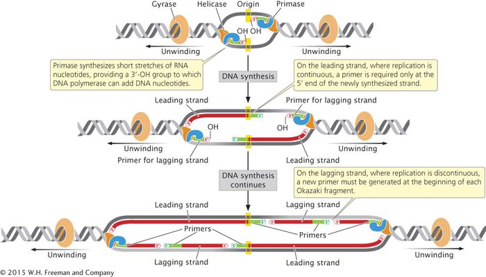
THE SYNTHESIS OF PRIMERS All DNA polymerases require a nucleotide with a 3′-OH group to which a new nucleotide can be added. Because of this requirement, DNA polymerases cannot initiate DNA synthesis on a bare template; rather, they require an existing 3′-OH group to get started. How, then, does DNA synthesis begin?
An enzyme called primase synthesizes short stretches (about 10–12 nucleotides long) of RNA nucleotides, or primers, which provide a 3′-OH group to which DNA polymerases can attach DNA nucleotides. (Because primase is an RNA polymerase, it does not require a preexisting 3′-OH group to start the synthesis of a nucleotide strand.) All DNA molecules initially have short RNA primers embedded within them; these primers are later removed and replaced with DNA nucleotides.
On the leading strand, where DNA synthesis is continuous, a primer is required only at the 5′ end of the newly synthesized strand. On the lagging strand, where replication is discontinuous, a new primer must be generated at the beginning of each Okazaki fragment (Figure 9.11). Primase forms a complex with helicase at the replication fork and moves along the template of the lagging strand. The single primer on the leading strand is probably synthesized by the primase–helicase complex on the template of the lagging strand of the other replication fork, at the opposite end of the replication bubble.
CONCEPTS
Primase synthesizes a short stretch of RNA nucleotides (a primer), which provides a 3′-OH group for the attachment of DNA nucleotides to start DNA synthesis.
 CONCEPT CHECK 4
Primers are synthesized where on the lagging strand?
Only at the 5′ end of the newly synthesized strand
Only at the 3′ end of the newly synthesized strand
At the beginning of every Okazaki fragment
At multiple places within an Okazaki fragment
c
DNA SYNTHESIS BY DNA POLYMERASES After DNA has unwound and a primer has been added, DNA polymerases elongate the polynucleotide strand by catalyzing DNA polymerization. The best-
| DNA polymerase | 5′→ 3′ polymerase activity | 3′→ 5′ exonuclease activity | 5′→ 3′ exonuclease activity | Function |
|---|---|---|---|---|
| I | Yes | Yes | Yes | Removes and replaces primers |
| III | Yes | Yes | No | Elongates DNA |
|
Note: DNA polymerases II, IV, and V are involved in DNA repair and translesion synthesis. |
||||
DNA polymerase III is a large multiprotein complex that acts as the main workhorse of replication. DNA polymerase III synthesizes nucleotide strands by adding new nucleotides to the 3′ end of a growing DNA strand. This enzyme has two enzymatic activities (see Table 9.2). Its 5′→3′ polymerase activity allows it to add new nucleotides in the 5′→3′ direction. Its 3′→5′ exonuclease activity allows it to remove nucleotides in the 3′→5′ direction, enabling it to correct errors. If a nucleotide with an incorrect base is inserted into the growing DNA strand, DNA polymerase III uses its 3′→5′ exonuclease activity to back up and remove the incorrect nucleotide. It then resumes its 5′→3′ polymerase activity. These two functions together allow DNA polymerase III to efficiently and accurately synthesize new DNA molecules.
The first E. coli polymerase to be discovered, DNA polymerase I, also has 5′→3′ polymerase and 3′→5′ exonuclease activities (see Table 9.2), which allow the enzyme to synthesize DNA and to correct errors. Unlike DNA polymerase III, however, DNA polymerase I also possesses 5′→3′ exonuclease activity, which is used to remove the primers laid down by primase and replace them with DNA nucleotides by synthesizing in a 5′→3′ direction (see Figure 9.12). The removal and replacement of primers appears to constitute the main function of DNA polymerase I.
Despite their differences, all of E. coli’s DNA polymerases
synthesize any sequence specified by the template strand.
synthesize in the 5′→3′ direction by adding nucleotides to a 3′-OH group.
use dNTPs to synthesize new DNA.
require a 3′-OH group to initiate synthesis.
catalyze the formation of a phosphodiester bond by joining the 5′-phosphate group of the incoming nucleotide to the 3′-OH group of the preceding nucleotide on the growing strand, cleaving off two phosphates in the process.
produce newly synthesized strands that are complementary and antiparallel to the template strands.
are associated with a number of other proteins.
 TRY PROBLEM 23
CONCEPTS
DNA polymerases synthesize DNA in the 5′→3′ direction by adding new nucleotides to the 3′ end of a growing nucleotide strand.
DNA LIGASE After DNA polymerase III attaches a DNA nucleotide to the 3′-OH group on the last nucleotide of the RNA primer, each new DNA nucleotide then provides the 3′-OH group needed for the next DNA nucleotide to be added. This process continues as long as template is available (Figure 9.12a). DNA polymerase I follows DNA polymerase III and, using its 5′→3′ exonuclease activity, removes the RNA primer. It then uses its 5′→3′ polymerase activity to replace the RNA nucleotides with DNA nucleotides. DNA polymerase I attaches the first nucleotide to the OH group at the 3′ end of the preceding Okazaki fragment and then continues, in the 5′→3′ direction along the nucleotide strand, removing and replacing, one at a time, the RNA nucleotides of the primer (Figure 9.12b).
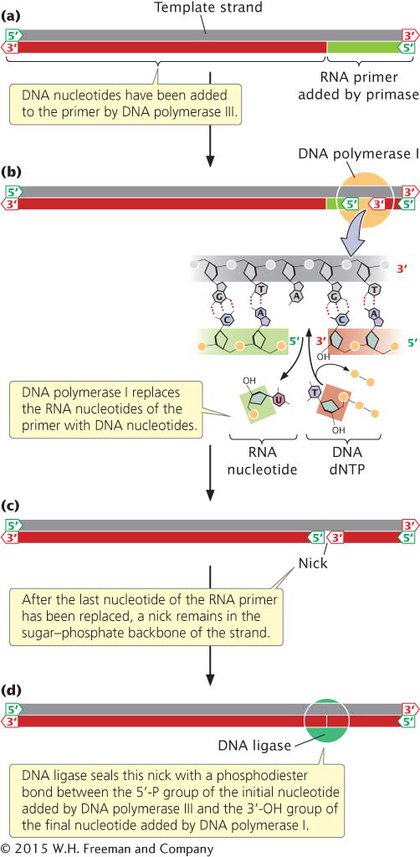
After polymerase I has replaced the last nucleotide of the RNA primer with a DNA nucleotide, a break remains in the sugar–phosphate backbone of the new DNA strand. The 3′-OH group of the last nucleotide to have been added by DNA polymerase I is not attached to the 5′-phosphate group of the first nucleotide added by DNA polymerase III (Figure 9.12c). This break is sealed by the enzyme DNA ligase, which catalyzes the formation of a phosphodiester bond without adding another nucleotide to the strand (Figure 9.12d). Some of the major enzymes and proteins required for prokaryotic DNA replication are summarized in Table 9.3.
| Component | Function |
|---|---|
| Initiator proteins | Bind to origin and separate strands of DNA to initiate replication |
| DNA helicase | Unwinds DNA at replication fork |
| Single- |
Attach to single- |
| DNA gyrase | Moves ahead of the replication fork, making and resealing breaks in the double- |
| DNA primase | Synthesizes a short RNA primer to provide a 3′-OH group for the attachment of DNA nucleotides |
| DNA polymerase III | Elongates a new nucleotide strand from the 3′-OH group provided by the primer |
| DNA polymerase I | Removes RNA primers and replaces them with DNA |
| DNA ligase | Joins Okazaki fragments by sealing breaks in the sugar–phosphate backbone of newly synthesized DNA |
CONCEPTS
After primers have been removed and replaced, the break in the sugar–phosphate linkage is sealed by DNA ligase.
CONCEPT CHECK 5
Which bacterial enzyme removes the primers?
Primase
DNA polymerase I
DNA polymerase III
Ligase
b
ELONGATION AT THE REPLICATION FORK Now that the major enzymatic components of elongation—
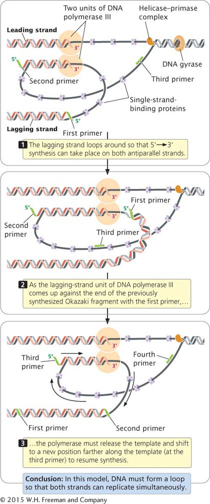
In summary, each active replication fork requires five basic components:
Helicase to unwind the DNA
Single-
strand- binding proteins to protect the single nucleotide strands and prevent secondary structures The topoisomerase gyrase to remove strain ahead of the replication fork
Primase to synthesize primers with a 3′-OH group at the beginning of each DNA fragment
DNA polymerase to synthesize the leading and lagging nucleotide strands
You can see how the different components of the replication process work together by viewing Animations 9.3 and 9.4.