CONCEPT38.5 Extraembryonic Membranes Protect and Nourish the Embryo
A major step in the evolution of tetrapods (four-limbed vertebrates) was the formation of the amniotic egg, seen today in mammals and reptiles, including birds. We saw in Concept 23.6 the significance of this egg. It provides the embryo with a contained aqueous environment, freeing the processes of reproduction and development from dependence on an external water supply. The three germ layers that form the embryo also create the membranes that provide this protective environment.
Extraembryonic membranes form with contributions from all germ layers
Whether they develop inside or outside the mother’s body, embryos of amniotes are surrounded by four extraembryonic membranes that function in protection, nutrition, gas exchange, and waste removal. The chicken embryo provides a good example, but the process is similar in other reptiles and in egg-laying mammals. (We will discuss placental mammals in the next section.) The four membranes are the yolk sac, allantois, amnion, and chorion (FIGURE 38.18).
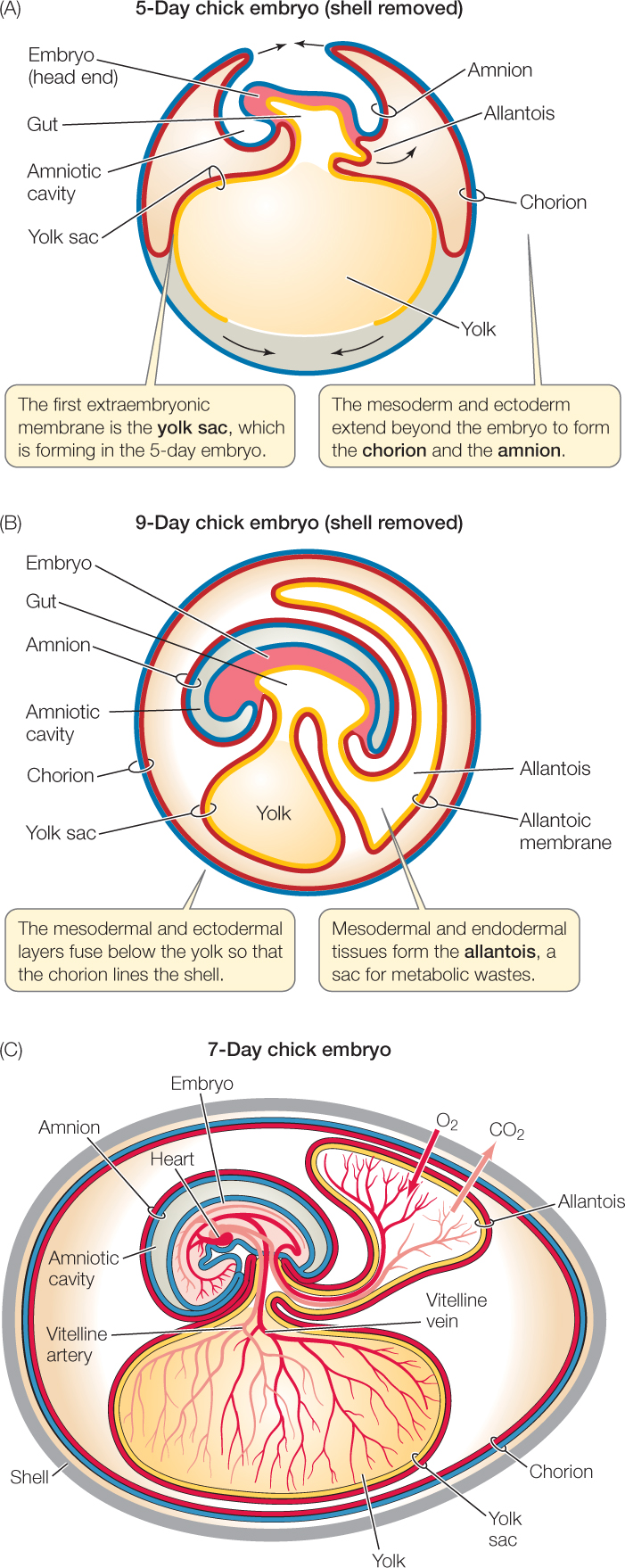
- The yolk sac forms first, by extension of the endoderm and the inner layer of lateral plate mesoderm (see Figure 38.18A,B). These two layers grow around the yolk and enclose it. The inner layer of lateral plate mesoderm is special because it can form blood cells and blood vessels. The first blood cells of the embryo and the vessels that carry them—the vitelline artery and its branches—form in the mesoderm of the yolk sac. Stored yolk is not transferred directly to the gut of the embryo through the yolk stalk. Rather, it is digested by the endoderm of the yolk sac, and the products are transported to the embryo by the developing blood and blood vessels in the yolk sac wall (Figure 38.18C).
- The allantois, a sac for storage of metabolic wastes, is also an outgrowth of endoderm and the inner layer of lateral plate mesoderm (Figure 38.18A–C). Because the yolk sac and the allantois are the extraembyronic membranes that contain lateral plate mesoderm, they are the only two that develop blood vessels (Figure 38.18C).
- Ectoderm and the outer layer of the lateral plate mesoderm also grow out from the embryo and surround it (Figure 38.18A). Where they meet, they fuse, forming two membranes, an inner amnion and outer chorion (Figure 38.18B). The amnion surrounds the embryo and secretes fluid into the enclosed cavity, providing a protective, shock-absorbing environment.
- The chorion, located just beneath the egg’s shell, forms a continuous membrane that limits water loss. The chorion cannot make blood vessels, but it fuses with the blood vessel–forming allantoic membrane, forming the chorioallantoic membrane that provides for exchange of O2 and CO2 between the embryo and the outside world (Figure 38.18C).
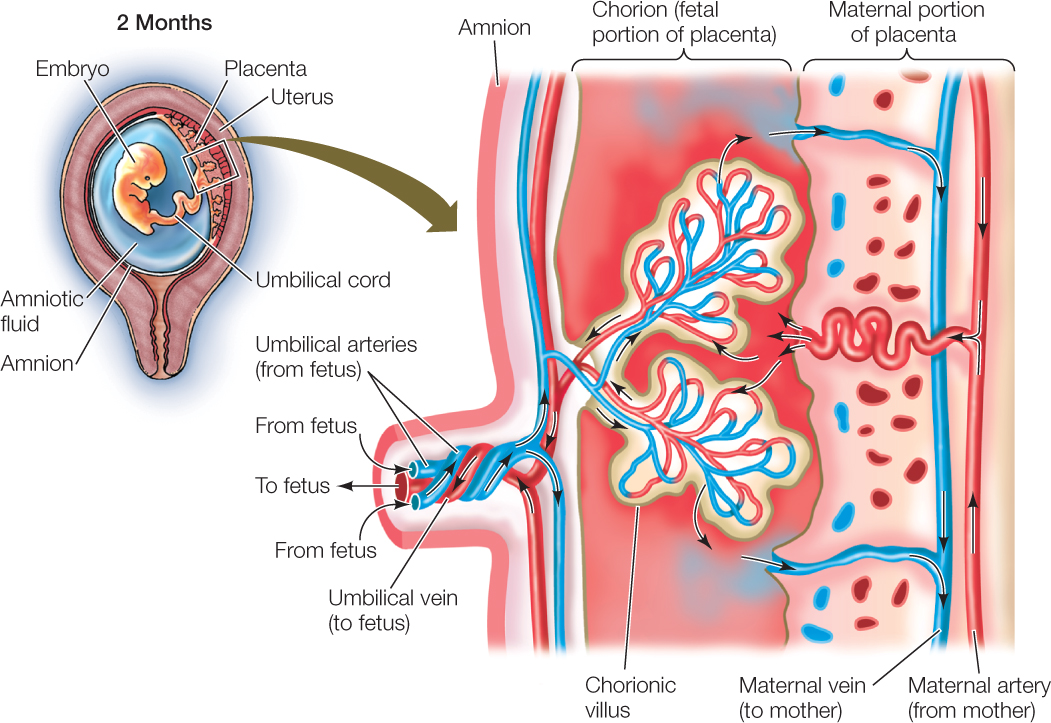
Extraembryonic membranes in mammals form the placenta
In placental mammals, the entire trophoblast becomes embedded in the endometrium of the uterus (see Figure 38.8). Hypoblast cells proliferate to form what in birds would be the yolk sac, even though there is little yolk in the eggs of placental mammals. In eutherian placental mammals, the allantois and chorion combine, forming the chorioallantoic placenta. These embryonic tissues, along with maternal tissue of the uterine wall (the endometrium), produce the placenta. Note that the placenta is a unique organ because it is composed of tissues from two organisms—the mother and her offspring. Interestingly, the yolk sac in mammals continues to be an early site of blood cell production as it is in birds, even though no or very little yolk is present. Meanwhile, the amnion grows around the embryo, enclosing it in a fluid-filled amniotic cavity (FIGURE 38.19). (A pregnant woman’s “water breaks” when the amnion bursts during labor and releases the amniotic fluid.)
Human gestation is traditionally divided into three periods of roughly 12 weeks each, called trimesters. The first trimester is a time of rapid cell division and tissue differentiation, and the embryo is very sensitive during this time to damage from radiation, drugs, chemicals, and pathogens that can cause birth defects such as those referred to in the opening story of the chapter. By the end of the first trimester, most organs have started to form, and the embryo is about 8 centimeters long. At about this time, the human embryo is medically and legally referred to as a fetus.
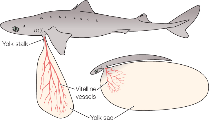
Fish also make yolk sacs
Many fish also have a very yolky egg (see Figure 38.7B). They are not amniotes, however, so how do they obtain nutrients from this stored material? Embryonic fish produce a yolk sac, but it differs from that of amniotes. As the fish embryo forms, all three germ layers—ectoderm, mesoderm and endoderm—grow around the yolk. The yolk sac becomes vascularized as in amniotes, and materials are carried in the blood vessels to the embryo (FIGURE 38.20).
804
CHECKpointCONCEPT38.5
- What are the four extraembryonic membranes of a chick embryo? Describe their functions.
- Would you expect a frog embryo to have the same extraembryonic membranes as a lizard? Why or why not?
- What function does the yolk sac have in both egg-laying and placental mammals?
- The yolk sac of fish is referred to as being trilaminar. Why is this term appropriate? How does a fish yolk sac differ from a chick yolk sac?
- What outstanding feature of the placenta makes it a unique organ?
We have now seen how a single-celled zygote becomes a complex organism, but development does not stop at birth or hatching. Animals must grow. For some, growth stops when they reach adulthood. Others grow throughout their lives. In either case, this growth is part of development. We will consider post-embryonic growth next.