THE PATH OF DIGESTION FROM MOUTH TO LARGE INTESTINE
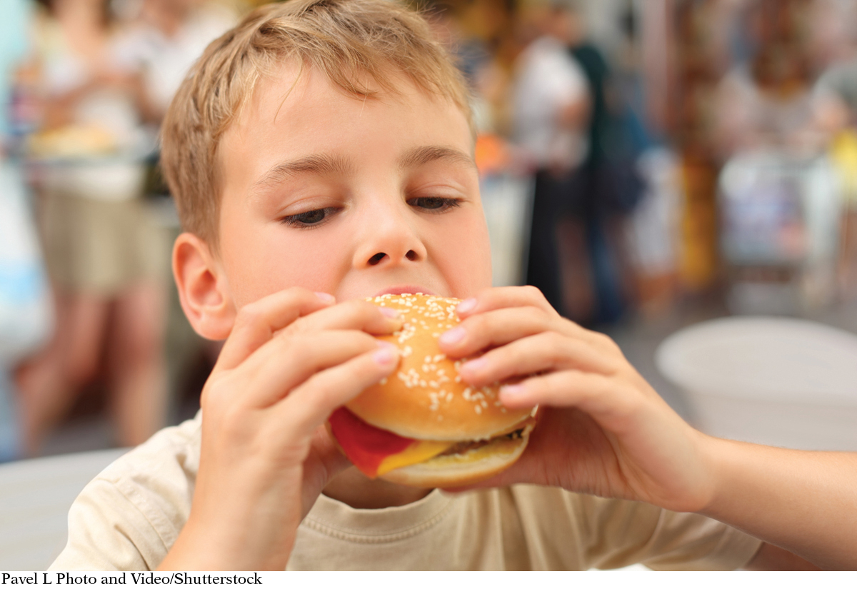
The object of digestion is to break down large molecules into smaller units, and the first stop is the mouth. Picture a hamburger poised to enter your mouth, or oral cavity. The first bite, or even the mouth-
SALIVA fluid secreted from salivary glands in the mouth to moisten food and provide lubrication
AMYLASES enzymes that break down starch into smaller polysaccharides and disaccharides
LIPASES enzymes that break down fats (triglycerides) by releasing one or more fatty acids
As you chew your mouthful of hamburger, the teeth tear and crush the bread and meat thus beginning mechanical digestion, which increases the surface area available for the enzymes to begin their work of chemical digestion. While we chew, the salivary glands near the jaw and under the tongue release saliva, which both lubricates the mouth and esophagus and contains salivary amylase, an enzyme that starts digesting the carbohydrates in the bun, as well as a lipase, which begins digesting fats in the meat. The tongue mixes saliva with the foods in the mouth and pushes food to the back of the mouth to initiate swallowing.
TASTE BUDS taste receptor cells found on the tongue within the papillae that are involved in sensing foods on the basis of specific flavors (tastes), such as sweet, sour, salty, bitter, and umami
The tongue does more than help mix and swallow the food, though—

These taste buds don’t just tell us if something is too bitter or salty; they also create signals that tell the rest of the GI tract to prepare for the next steps of digestion.
Food Passes from the Esophagus to the Stomach
BOLUS a masticated, round lump of food, lubricated in the mouth by mixing with saliva
SPHINCTER a ring-
Once the hamburger has been chewed and coated in saliva, it becomes a soft, moist lump of food known as a bolus, which is swallowed and passed through the throat. It then enters the esophagus, a roughly 10-
PROTEASES enzymes that break down proteins
CHYME semi-
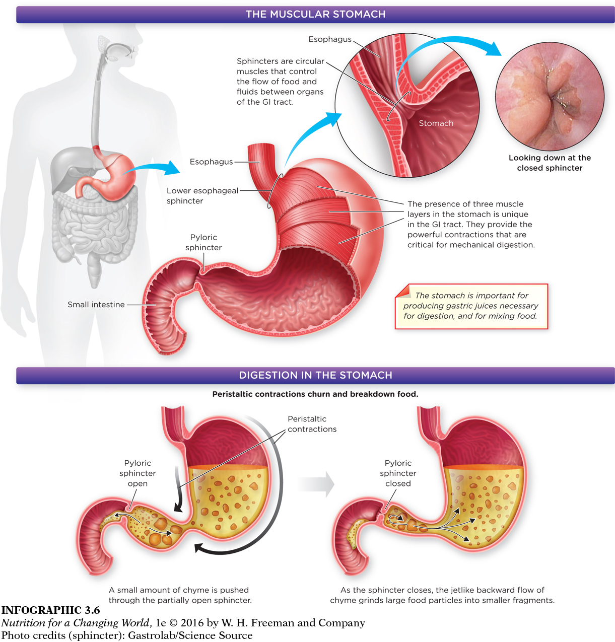
The bolus then enters the stomach, a muscular, J-
Question 3.6
 What structural feature is found in the stomach that is responsible for the strength of its muscle contractions?
The contractions of the stomach are strong because the stomach has three layers of muscles.
How quickly the stomach empties into the small intestine depends on the composition and quantity of the foods and fluids you consume. A hamburger typically spends 24 to 72 hours going from the mouth to the anus, but this transit time can change because of illness, medication, how active you are, and even your emotional state. Food with more fiber, for instance, slows emptying from the stomach, helping you feel full. But as fiber passes into the large intestine, it can also stimulate propulsive contractions, which speeds up the transit of the intestinal contents through the rest of the digestive system. These combined effects of fiber are important and help explain why nutrition professionals recommend healthy amounts of vegetables, fruits and other high-
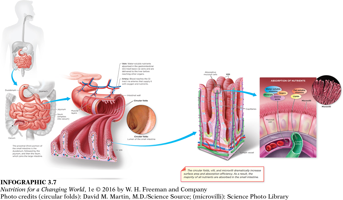
The Structural Features of the Small Intestine Facilitate Absorption of Nutrients
VILLI fingerlike projections that protrude from the absorptive mucosal cells of the small intestine into the lumen of the GI tract; responsible for increasing the available surface area for absorption
MICROVILLI very small projections that protrude from the absorptive mucosal cells of the villi in the small intestine; responsible for increasing the surface area for absorption twenty-
BRUSH BORDER name for the microvilli-
The small intestine is the primary site for the digestion of food and the absorption of nutrients and as we shall see, it is where digestion goes awry in those with celiac disease. The small intestine has three sections: the duodenum, the first portion of the small intestine after the stomach; the jejenum, the middle portion; and the ileum, the last and longest portion. The small intestine isn’t actually “small” at all; it is a coiled hollow tube approximately 20 feet long and one-
Question 3.7
List the three structural features of the small intestine that cause it to have a much larger surface area than a simple pipe of the same length would have.
The surface area of the small intestine is large because of the circular folds of the lumen of the small intestine, the villi that protrude from the mucosal cells of the small intestine, and the microvilli that protrude from the mucosal cells of the villi of the small intestine. These structures work together to optimize nutrient absorption.
Secretions from Accessory Organs Aid in Digestion
Secretions from accessory organs such as the pancreas and gallbladder play an important role in the digestion of the hamburger within the lumen of the small intestine. Chyme that enters the small intestine from the stomach is very acidic, and if it doesn’t get neutralized, it denatures and inactivates the enzymes required for digestion. The pancreas releases pancreatic juice that contains bicarbonate (baking soda), a base that neutralizes the gastric acids in chyme. Refer to INFOGRAPHIC 3.3.
BILE a fluid produced in the liver, concentrated and stored in the gallbladder, and secreted into the small intestine in response to food present in stomach; bile promotes the digestion of fat by emulsifying it, which allows lipase easier access
The liver and the gallbladder also help to digest the many lipids found in the hamburger. The liver produces bile—which is stored in the gallbladder, a small, pear-
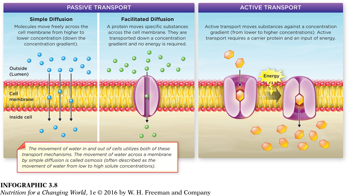
Transport of Nutrients across the Cell Membrane
SIMPLE DIFFUSION movement of a substance across a cell membrane, down a concentration gradient
FACILITATED DIFFUSION movement of a substance across a cell membrane, down a concentration gradient, with the assistance of a specific transport protein
ACTIVE TRANSPORT the energy-
To enter the mucosal cells lining the GI tract, water and small amounts of a few other nutrients can pass directly through the cell membrane by simple diffusion. The cell membrane serves as the boundary that holds the content of body’s cells in place and keeps their internal structures safe, so that cells function properly. The membrane also serves as a semi-
Question 3.8
What is the only transport mechanism that can transport substances against a concentration gradient?
Active transport is the only transport mechanism that can move substances against a concentration gradient (from an area of lower concentration to higher concentration). This form of transport requires energy.
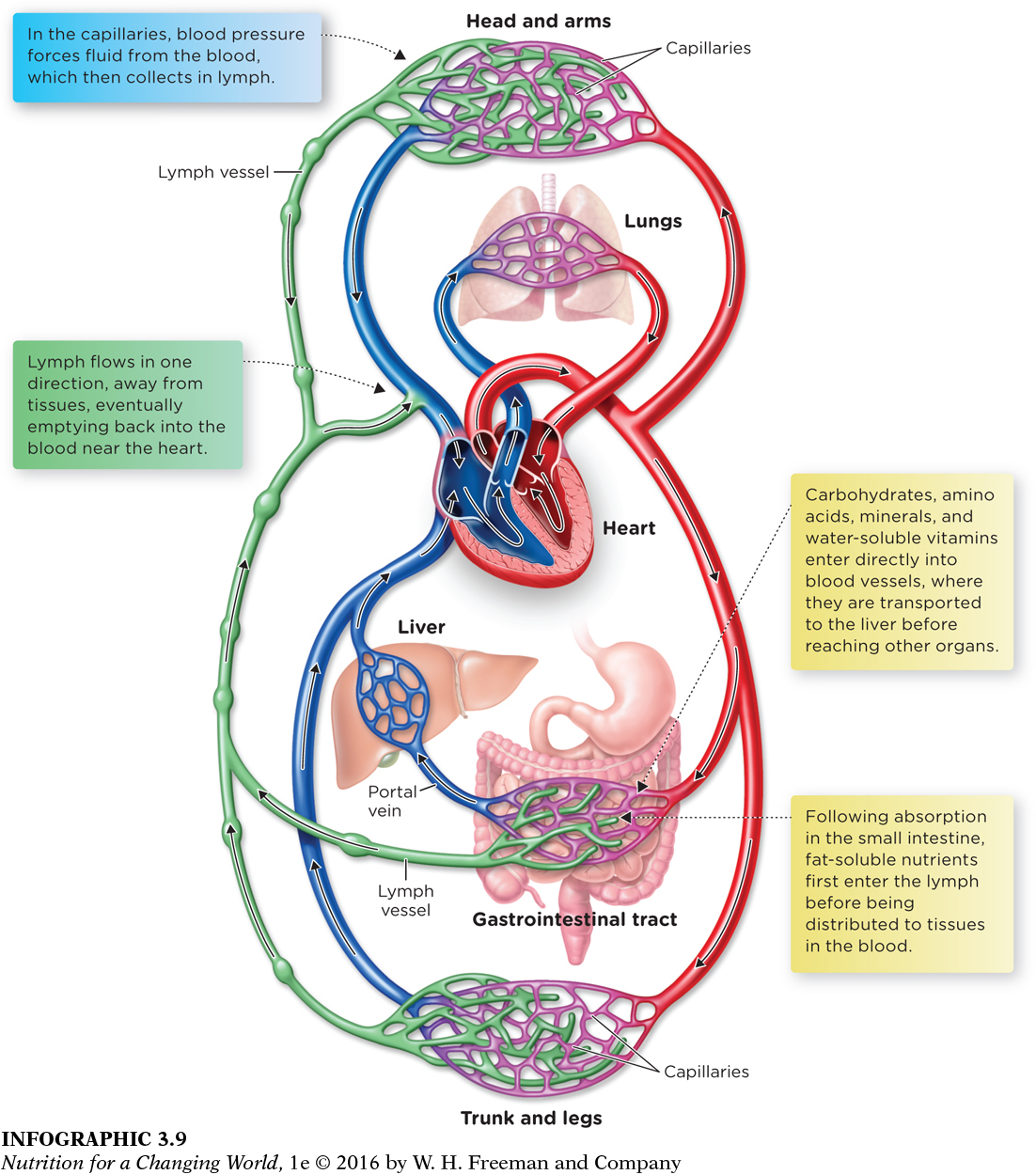
Circulation of Nutrients in Blood and Lymph
CIRCULATORY SYSTEM a system made up of veins, arteries, capillaries, heart and lymphatic vessels; responsible for movement of blood and lymph throughout the body
LYMPHATIC SYSTEM a system of vessels in which the products of fat digestion, among other things, are transported from the GI tract to the blood
Once inside the cells of the brush border of the small intestine, nutrients must reach the areas of the body where they are needed. The circulatory system which includes both the blood and the lymphatic system distribute vital nutrients to the tissues and organs. For example, carbohydrates, amino acids, minerals, and water-
Question 3.9
How do lipids that leave the small intestine in lymph get to tissues throughout the body?
Lipids leave the small intestine in lymph and flow toward the heart where the lymph empties into the bloodstream. The heart then pumps the lipids to tissues throughout the body.
■ ■ ■
For people with celiac disease, however, these last few steps do not progress properly, and the delicate and essential process of absorption becomes disrupted. The gluten found in the hamburger bun sparks an immune reaction that triggers the person’s immune cells to attack other body cells. These so-
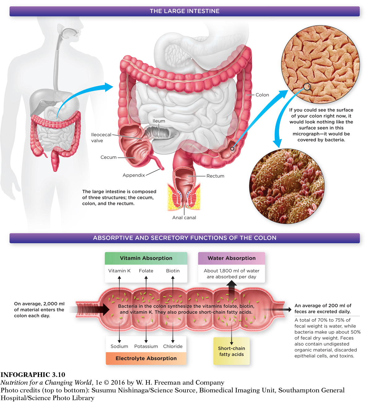
The Large Intestine is the End of the Gastrointestinal Tract
From the small intestine, any undigested nutrients that remain in the chyme are passed into the large intestine, which consists of the cecum, colon, and the rectum. Here, little digestion or absorption take place; there are no villi. Secreted mucus protects and lubricates the lining, making it easier for everything that’s leftover to be excreted as feces. (INFOGRAPHIC 3.10)
Question 3.10
What is the name of the structure that prevents the movement of fecal material into the small intestine?
The ileocecal valve prevents fecal material from entering the small intestine.
But before that happens, the large intestine will extract electrolytes (sodium, chloride, and potassium), some fatty acids, vitamins (K, biotin, and folate), and water. In addition, the large intestine contains more than 1,000 species of bacteria that feed on undigested fiber and starch; since humans have no enzymes that digest dietary fiber, these bacteria perform some of that function, producing gas and short-