3.4 Structure of the Brain
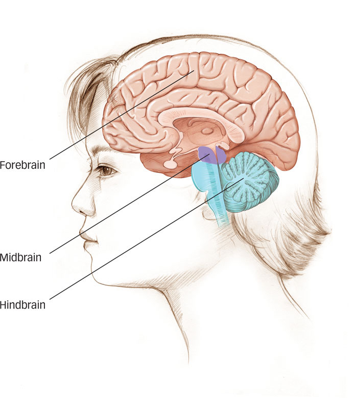
The human brain, weighing in at about three pounds, is really not much to look at. You already know that its neurons and glial cells are busy humming away, giving you potentially brilliant ideas, consciousness, and feelings. But which neurons in which parts of the brain control which functions? To answer that question, neuroscientists had to find a way of describing the brain that allows researchers to communicate with one another. It can be helpful to talk about areas of the brain from “bottom to top,” noting how the different regions are specialized for different kinds of tasks. In general, simpler functions are performed at the “lower levels” of the brain, whereas more complex functions are performed at successively “higher” levels (see FIGURE 3.13). Or, as you’ll see shortly, the brain can also be approached in a “side-
96
Let’s look first at the divisions of the brain, and the responsibilities of each part, moving from the bottom to the top. Using this view, we can divide the brain into three parts: the hindbrain, the midbrain, and the forebrain (see Figure 3.13).
The Hindbrain

hindbrain An area of the brain that coordinates information coming into and out of the spinal cord.
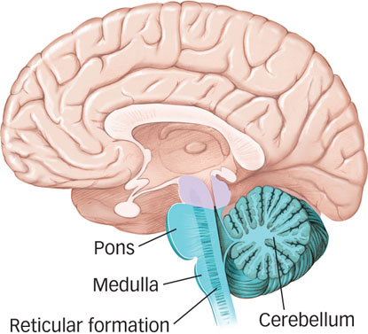
If you follow the spinal cord from your tailbone to where it enters your skull, you’ll find it difficult to determine where your spinal cord ends and your brain begins. That’s because the spinal cord is continuous with the hindbrain, an area of the brain that coordinates information coming into and out of the spinal cord. The hindbrain looks like a stalk on which the rest of the brain sits, and it controls the most basic functions of life: respiration, alertness, and motor skills. The structures that make up the hindbrain include: the medulla, the reticular formation, the cerebellum, and the pons (see FIGURE 3.14).
medulla An extension of the spinal cord into the skull that coordinates heart rate, circulation, and respiration.
The medulla is an extension of the spinal cord into the skull that coordinates heart rate, circulation, and respiration. Beginning inside the medulla and extending upward is a small cluster of neurons called the reticular formation, which regulates sleep, wakefulness, and levels of arousal. In one early experiment, researchers stimulated the reticular formation of a sleeping cat. This caused the animal to awaken almost instantaneously and remain alert. Conversely, severing the connections between the reticular formation and the rest of the brain caused the animal to lapse into an irreversible coma (Moruzzi & Magoun, 1949). The reticular formation maintains the same delicate balance between alertness and unconsciousness in humans. In fact, many general anesthetics work by reducing activity in the reticular formation, rendering the patient unconscious.
reticular formation A brain structure that regulates sleep, wakefulness, and levels of arousal.
Which part of the brain helps to orchestrate movements that keep you steady on your bike?
Behind the medulla is the cerebellum, a large structure of the hindbrain that controls fine motor skills. (Cerebellum is Latin for “little brain,” and the structure does look like a small replica of the brain.) The cerebellum orchestrates the proper sequence of movements when we ride a bike, play the piano, or maintain balance while walking and running. It contributes to the fine-
cerebellum A large structure of the hindbrain that controls fine motor skills.
pons A brain structure that relays information from the cerebellum to the rest of the brain.
The last major area of the hindbrain is the pons, a structure that relays information from the cerebellum to the rest of the brain. (Pons means “bridge” in Latin.) Although the detailed functions of the pons remain poorly understood, it essentially acts as a relay station or bridge between the cerebellum and other structures in the brain.
The Midbrain
tectum A part of the midbrain that orients an organism in the environment.
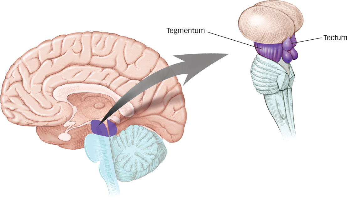
Sitting on top of the hindbrain is the midbrain, which is relatively small in humans. As you can see in FIGURE 3.15, the midbrain contains two main structures: the tectum and the tegmentum. The tectum orients an organism in the environment. The tectum receives stimulus input from the eyes, ears, and skin and moves the organism in a coordinated way toward the stimulus. For example, when you’re studying in a quiet room and you hear a click behind and to the right of you, your body will swivel and orient to the direction of the sound; this is your tectum in action.
97
tegmentum A part of the midbrain that is involved in movement and arousal.
The tegmentum is involved in movement and arousal; it also helps to orient an organism toward sensory stimuli. The midbrain may be relatively small, but it is a central location of neurotransmitters involved in arousal, mood, and motivation and the brain structures that rely on them (White, 1996). You could survive if you had only a hindbrain and a midbrain. The structures in the hindbrain would take care of all the bodily functions necessary to sustain life, and the structures in the midbrain would orient you toward or away from pleasurable or threatening stimuli in the environment. But this wouldn’t be much of a life. To understand where the abilities that make us fully human come from, we need to consider the last division of the brain.
The Forebrain
When you appreciate the beauty of a poem, detect the sarcasm in a friend’s remark, plan to go skiing next winter, or notice the faint glimmer of sadness on a loved one’s face, you are enlisting the forebrain. The forebrain is the highest level of the brain—
cerebral cortex The outermost layer of the brain, visible to the naked eye and divided into two hemispheres.
The cerebral cortex is the outermost layer of the brain, visible to the naked eye, and divided into two hemispheres. The subcortical structures are areas of the forebrain housed under the cerebral cortex near the center of the brain (see FIGURE 3.16). We’ll have much more to say about the two hemispheres of the cerebral cortex and the functions they serve in the next section, fittingly saving the highest level of the brain for last. First, we’ll examine the subcortical structures.
subcortical structures Areas of the forebrain housed under the cerebral cortex near the very center of the brain.
Subcortical Structures
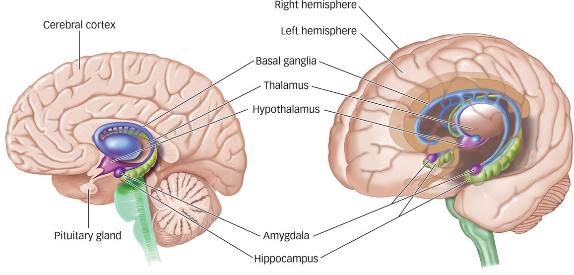
The subcortical (beneath the cortex) structures are nestled deep inside the brain, where they are quite protected. If you imagine sticking an index finger in each of your ears and pushing inward until they touch, that’s about where you’d find the thalamus, hypothalamus, pituitary gland, limbic system, and basal ganglia (see Figure 3.16). Each of these subcortical structures plays an important role in relaying information throughout the brain, as well as performing specific tasks that allow us to think, feel, and behave as humans. Here we’ll give you a brief introduction to each, and you’ll read more about many of these structures in later chapters.
98
Thalamus, Hypothalamus, and Pituitary Gland. The thalamus, hypothalamus, and pituitary gland, located in the center of the brain, interact closely with several other brain structures. They relay signals to and from these structures and also help to regulate them.
How is the thalamus like a computer?
thalamus A subcortical structure that relays and filters information from the senses and transmits the information to the cerebral cortex.
The thalamus relays and filters information from the senses and transmits the information to the cerebral cortex. The thalamus receives inputs from all the major senses except smell, which has direct connections to the cerebral cortex. The thalamus acts as a kind of computer server in a networked system, taking in multiple inputs and relaying them to a variety of locations (Guillery & Sherman, 2002). However, unlike the mechanical operations of a computer (“send input A to location B”), the thalamus actively filters sensory information, giving more weight to some inputs and less weight to others. The thalamus also closes the pathways of incoming sensations during sleep, providing a valuable function in not allowing information to pass to the rest of the brain.

hypothalamus A subcortical structure that regulates body temperature, hunger, thirst, and sexual behavior.
The hypothalamus, located below the thalamus (hypo-
99
pituitary gland The “master gland” of the body’s hormone-
Located below the hypothalamus is the pituitary gland, the “master gland” of the body’s hormone-
limbic system A group of forebrain structures including the hypothalamus, the hippocampus, and the amygdala, which are involved in motivation, emotion, learning, and memory.
The Limbic System. The hypothalamus also is part of the limbic system, a group of forebrain structures including the hypothalamus, the hippocampus, and the amygdala, which are involved in motivation, emotion, learning, and memory (Maclean, 1970; Papez, 1937). The limbic system is where the subcortical structures meet the cerebral cortex.
hippocampus A structure critical for creating new memories and integrating them into a network of knowledge so that they can be stored indefinitely in other parts of the cerebral cortex.
The hippocampus (from Latin for “sea horse,” due to its shape) is critical for creating new memories and integrating them into a network of knowledge so that they can be stored indefinitely in other parts of the cerebral cortex. Individuals with damage to the hippocampus can acquire new information and keep it in awareness for a few seconds, but as soon as they are distracted, they forget the information and the experience that produced it (Scoville & Milner, 1957; Squire, 2009). This kind of disruption is limited to everyday memory for facts and events that we can bring to consciousness; memory of learned habitual routines or emotional reactions remains intact (Squire, Knowlton, & Musen, 1993). As an example, people with damage to the hippocampus can remember how to drive and talk, but they cannot recall where they have recently driven or a conversation they have just had. You will read more about the hippocampus and its role in creating, storing, and combining memories in the Memory chapter.
Why are you likely to remember details of a traumatic event?

amygdala A part of the limbic system that plays a central role in many emotional processes, particularly the formation of emotional memories.
The amygdala (from Latin for “almond,” also due to its shape), located at the tip of each horn of the hippocampus, plays a central role in many emotional processes, particularly the formation of emotional memories (Aggleton, 1992). The amygdala attaches significance to previously neutral events that are associated with fear, punishment, or reward (LeDoux, 1992). As an example, think of the last time something scary or unpleasant happened to you: A car came barreling toward you as you started walking into an intersection or a ferocious dog leaped out of an alley as you passed by. Those stimuli—
100
basal ganglia A set of subcortical structures that directs intentional movements.
The Basal Ganglia. There are several other structures in the subcortical area, but we’ll consider just one more. The basal ganglia are a set of subcortical structures that directs intentional movements. The basal ganglia are located near the thalamus and hypothalamus; they receive input from the cerebral cortex and send outputs to the motor centers in the brain stem. One part of the basal ganglia, the striatum, is involved in the control of posture and movement. As we saw in the excerpt from Michael J. Fox’s book, people who suffer from Parkinson’s disease typically show symptoms of uncontrollable shaking and sudden jerks of the limbs and are unable to initiate a sequence of movements to achieve a specific goal. This happens because the dopamine-
So, what’s the problem in Parkinson’s: the jerky movements, the ineffectiveness of the striatum in directing behavior, the botched interplay of the substantia nigra and the striatum, or the underproduction of dopamine at the neuronal level? The answer is all of the above. This unfortunate disease provides a nice illustration of two themes regarding the brain and behavior. First, invisible actions at the level of neurons in the brain can produce substantial effects at the level of behavior. Second, the interaction of hindbrain, midbrain, and forebrain structures shows how the various regions are interdependent.
The Cerebral Cortex
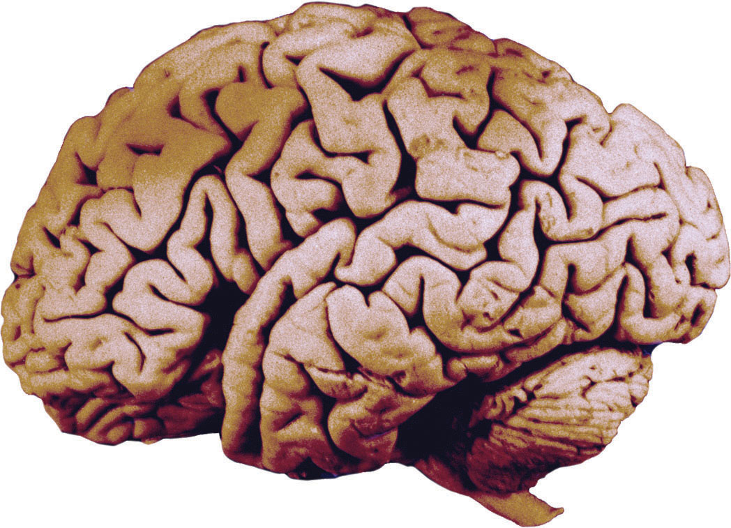
Our tour of the brain has taken us from the very small (neurons) to the somewhat bigger (major divisions of the brain) to the very large: the cerebral cortex. The cortex is the highest level of the brain, and it is responsible for the most complex aspects of perception, emotion, movement, and thought (Fuster, 2003). It sits over the rest of the brain, like a mushroom cap shielding the underside and stem, and it is the wrinkled surface you see when looking at the brain with the naked eye.
The smooth surfaces of the cortex—
Organization across Hemispheres. The first level of organization divides the cortex into the left and right hemispheres. The two hemispheres are more or less symmetrical in their appearance and, to some extent, in their functions. However, each hemisphere controls the functions of the opposite side of the body. This is called contralateral control, meaning that your right cerebral hemisphere perceives stimuli from and controls movements on the left side of your body, whereas your left cerebral hemisphere perceives stimuli from and controls movement on the right side of your body.
101
corpus callosum A thick band of nerve fibers that connects large areas of the cerebral cortex on each side of the brain and supports communication of information across the hemispheres.
The cerebral hemispheres are connected to each other by commissures, bundles of axons that make possible communication between parallel areas of the cortex in each half. The largest of these commissures is the corpus callosum, which connects large areas of the cerebral cortex on each side of the brain and supports communication of information across the hemispheres (see FIGURE 3.18). This means that information received in the right hemisphere, for example, can pass across the corpus callosum and be registered, virtually instantaneously, in the left hemisphere.
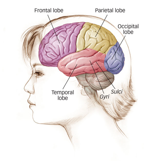
Organization within Hemispheres. The second level of organization in the cerebral cortex distinguishes the functions of the different regions within each hemisphere of the brain. Each hemisphere of the cerebral cortex is divided into four areas, or lobes: From back to front, these are the occipital lobe, the parietal lobe, the temporal lobe, and the frontal lobe, as shown in Figure 3.17. We’ll examine the functions of these lobes in more detail in later chapters, noting how scientists have used a variety of techniques to understand the operations of the brain. For now, here’s a brief overview of the main functions of each lobe.
occipital lobe A region of the cerebral cortex that processes visual information.
The occipital lobe, located at the back of the cerebral cortex, processes visual information. Sensory receptors in the eyes send information to the thalamus, which in turn sends information to the primary areas of the occipital lobe, where simple features of the stimulus are extracted, such as the location and orientation of an object’s edges (see the Sensation and Perception chapter for more details). These features are then processed into a more complex “map” of the stimulus onto the occipital cortex, leading to comprehension of what’s being seen. As you might imagine, damage to the primary visual areas of the occipital lobe can leave a person with partial or complete blindness. Information still enters the eyes, which work just fine. But without the ability to process and make sense of the information at the level of the cerebral cortex, the information is as good as lost (Zeki, 2001).
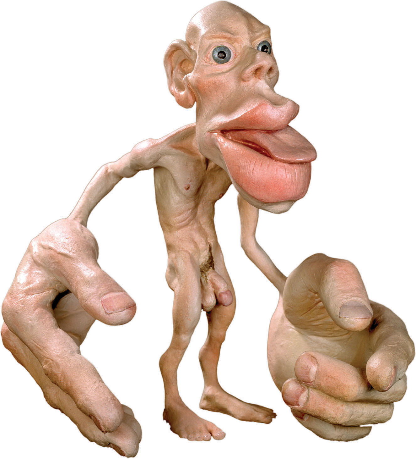
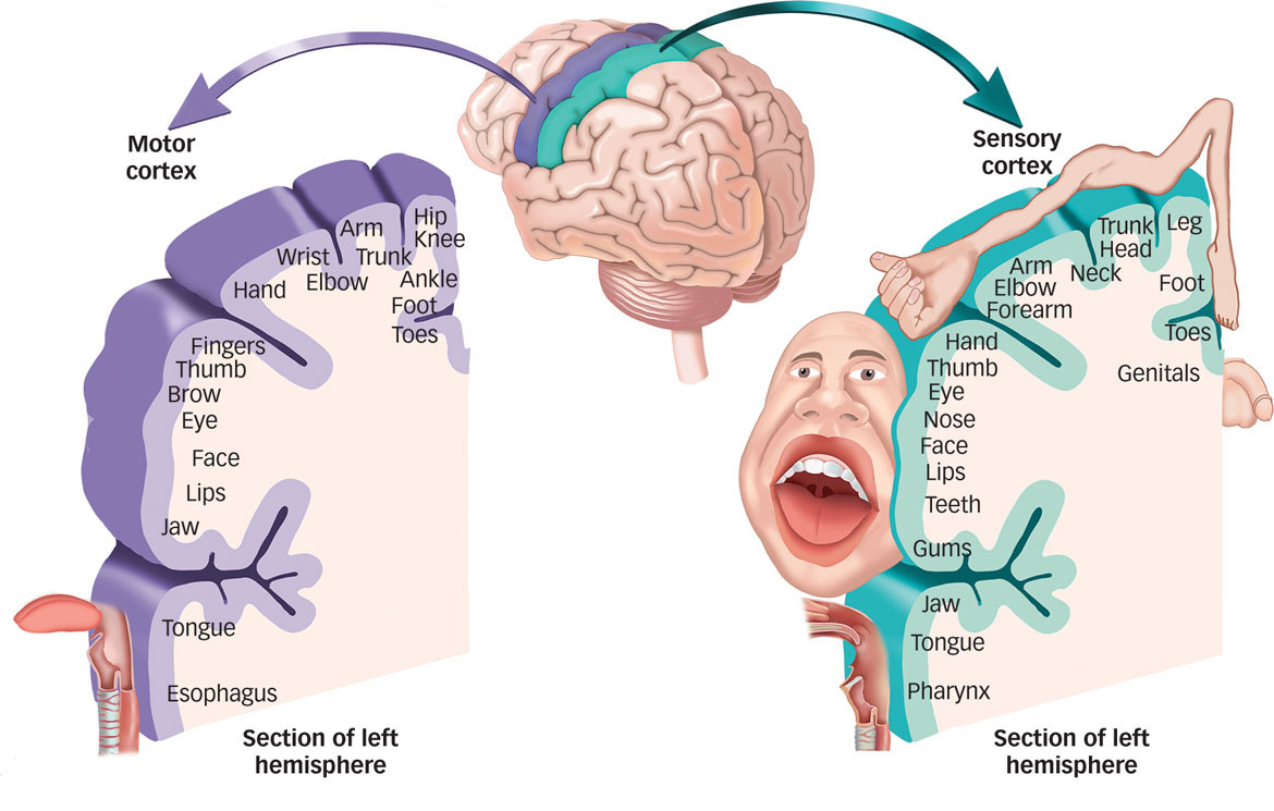
Why is the part of the somatosensory cortex relating to the lips bigger than the area corresponding to the feet?
parietal lobe A region of the cerebral cortex whose functions include processing information about touch.
The parietal lobe, located in front of the occipital lobe, carries out functions that include -processing information about touch. The parietal lobe contains the somatosensory cortex, a strip of brain tissue running from the top of the brain down to the sides (see FIGURE 3.19). Within each hemisphere, the somatosensory cortex represents the skin areas on the contralateral surface of the body. Each part of the somatosensory cortex maps onto a particular part of the body. If a body area is more sensitive, a larger part of the somatosensory cortex is devoted to it. For example, the part of the somatosensory cortex that corresponds to the lips and tongue is larger than the area corresponding to the feet. The somatosensory cortex can be illustrated as a distorted figure, called a homunculus (“little man”), in which the body parts are rendered according to how much of the somatosensory cortex is devoted to them (Penfield & Rasmussen, 1950). Directly in front of the somatosensory cortex, in the frontal lobe, is a parallel strip of brain tissue called the motor cortex. Like the somatosensory cortex, different parts of the motor cortex correspond to different body parts. The motor cortex initiates voluntary movements and sends messages to the basal ganglia, cerebellum, and spinal cord. The motor and somatosensory cortices, then, are like sending and receiving areas of the cerebral cortex, taking in information and sending out commands as the case might be.
102
temporal lobe A region of the cerebral cortex responsible for hearing and language.
The temporal lobe, located on the lower side of each hemisphere, is responsible for hearing and language. The primary auditory cortex in the temporal lobe is analogous to the somatosensory cortex in the parietal lobe and the primary visual areas of the occipital lobe: It receives sensory information from the ears based on the frequencies of sounds (Recanzone & Sutter, 2008). Secondary areas of the temporal lobe then process the information into meaningful units, such as speech and words. The temporal lobe also houses the visual association areas that interpret the meaning of visual stimuli and help us recognize common objects in the environment (Martin, 2007).
What types of thinking occur in the frontal lobe?
frontal lobe A region of the cerebral cortex that has specialized areas for movement, abstract thinking, planning, memory, and judgment.
The frontal lobe, which sits behind the forehead, has specialized areas for movement, abstract thinking, planning, memory, and judgment. As you just read, it contains the motor cortex, which coordinates movements of muscle groups throughout the body. Other areas in the frontal lobe coordinate thought processes that help us manipulate information and retrieve memories, which we can use to plan our behaviors and interact socially with others. In short, the frontal cortex allows us to do the kind of thinking, imagining, planning, and anticipating that sets humans apart from most other species (Schoenemann, Sheenan, & Glotzer, 2005; Stuss & Benson, 1986; Suddendorf & Corballis, 2007).
association areas Areas of the cerebral cortex that are composed of neurons that help provide sense and meaning to information registered in the cortex.
Organization within Specific Lobes. The third level of organization in the cerebral cortex involves the representation of information within specific lobes in the cortex. There is a hierarchy of processing stages from primary areas that handle fine details of information all the way up to association areas, which are composed of neurons that help provide sense and meaning to information registered in the cortex. For example, neurons in the primary visual cortex are highly specialized: some detect features of the environment that are in a horizontal orientation, others detect movement, and still others process information about human versus nonhuman forms. Secondary areas interpret the information extracted by these primary areas (shape, motion, etc.) to make sense of what’s being perceived; in this case, perhaps a large cat leaping toward your face. Similarly, neurons in the primary auditory cortex register sound frequencies, but it’s the association areas of the temporal lobe that allow you to turn those noises into the meaning of your friend screaming, “Look out for the cat!” Association areas, then, help stitch together the threads of information in the various parts of the cortex to produce a meaningful understanding of what’s being registered in the brain.
103

mirror neurons Neurons that are active when an animal performs a behavior, such as reaching for or manipulating an object, and are also activated when another animal observes that animal performing the same behavior.
A striking example of this property of association areas comes from the discovery of the mirror-
Finally, neurons in the association areas are usually less specialized and more flexible than neurons in the primary areas. As such, they can be shaped by learning and experience to do their job more effectively. This kind of shaping of neurons by environmental forces allows the brain flexibility, or plasticity, our next topic.
Brain Plasticity
What does it mean to say that the brain is plastic?
The cerebral cortex may seem like a fixed structure, one big sheet of neurons designed to help us make sense of our external world. Remarkably, though, sensory cortices are not fixed. They can adapt to changes in sensory inputs, a quality researchers call plasticity (i.e., the ability to be molded). As an example, if you lose your middle finger in an accident, the part of the somatosensory area that represents that finger is initially unresponsive (Kaas, 1991). After all, there’s no longer any sensory input going from that location to that part of the brain. You might expect the left middle-
Plasticity doesn’t only occur to compensate for missing digits or limbs, however. An extraordinary amount of stimulation of one finger can result in that finger “taking over” the representation of the part of the cortex that usually represents other, adjacent fingers (Merzenich et al., 1990). For example, concert pianists have highly developed cortical areas for finger control: The continued input from the fingers commands a larger area of representation in the somatosensory cortices in the brain. Consistent with this observation, recent research indicates greater plasticity within the motor cortex of professional musicians compared with nonmusicians, perhaps reflecting an increase in the number of motor synapses as a result of extended practice (Rosenkranz, Williamon, & Rothwell, 2007). Similar findings have been obtained with quilters (who have highly developed areas for the thumb and forefinger, which are critical to their profession) and taxi drivers (who have overdeveloped brain areas in the hippocampus that are used during spatial navigation; Maguire, Woollett, & Spiers, 2006).
104
THE REAL WORLD: Brain Plasticity and Sensations in Phantom Limbs
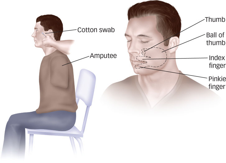
Long after a limb is amputated, many patients continue to experience sensations where the missing limb would be, a phenomenon called phantom limb syndrome. Patients can feel their missing limbs moving, even in coordinated gestures such as shaking hands. Some even report feeling pain in their phantom limbs. Why does this happen? Some evidence suggests that phantom limb syndrome may arise in part because of plasticity in the brain.
Researchers stimulated the skin surface in various regions around the face, torso, and arms while monitoring brain activity in amputees and non-
Brain scans of the amputees revealed that stimulating areas of the face and upper arm activated an area in the somatosensory cortex that previously would have been activated by a now-
Brain plasticity can explain these results (Pascual-
This and related research suggest one explanation for a previously poorly understood phenomenon. How can a person “feel” something that isn’t there? Brain plasticity, an adaptive process through which the brain reorganizes itself, offers an answer (Flor, Nikolajsen, & Jensen, 2006). The brain established new mappings that led to novel sensations.
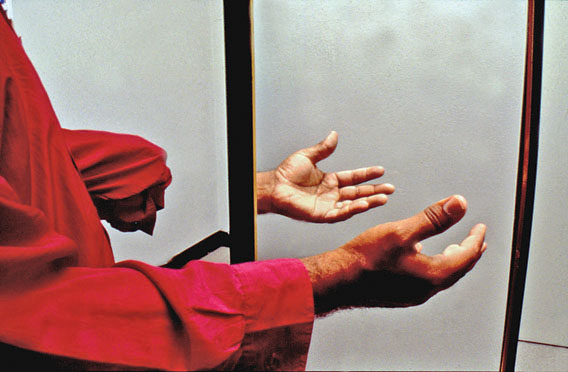
This idea also has practical implications for dealing with the pain that can result from phantom limbs (Ramachandran & Altschuler, 2009). Researchers have used a “mirror box” to teach patients a new mapping to increase voluntary control over their phantom limbs. For example, a patient would place his intact right hand and phantom left hand in the mirror box such that when looking at the mirror, he sees his right hand reflected on the left—
105
Plasticity is also related to a question you might not expect to find in a psychology text: How much exercise have you been getting lately? While we expect that you are spending countless happy hours reading this text, we also hope that you’ve been finding enough time for physical exercise. A large of number of studies in rats and other nonhuman animals indicate that physical exercise can increase the number of synapses and even promote the development of new neurons in the hippocampus (Hillman, Erickson, & Kramer, 2008; van Praag, 2009). Recent studies with people have begun to document beneficial effects of cardiovascular exercise on aspects of brain function and cognitive performance (Colcombe et al., 2004, 2006). Although these effects tend to be seen most clearly in older adults (okay, so it’s time for your textbook authors to get on a treadmill), benefits have also been documented throughout the life span (Hertig & Nagel, 2012; Hillman et al., 2008; Roig et al., 2012). In fact, some researchers believe that this kind of activity-
 The brain can be divided into the hindbrain, midbrain, and forebrain.
-
The hindbrain generally coordinates information coming into and out of the spinal cord with structures such as the medulla, the reticular formation, the cerebellum, and the pons. These structures respectively coordinate breathing and heart rate, regulate sleep and arousal levels, coordinate fine motor skills, and communicate this information to the cortex.
-
The structures of the midbrain, the tectum and tegmentum, generally coordinates functions such as orientation to the environment and movement and arousal toward sensory stimuli.
-
The forebrain generally coordinates higher-
level functions, such as perceiving, feeling, and thinking. The forebrain houses subcortical structures, such as the thalamus, hypothalamus, limbic system (including the hippocampus and amygdala), and basal ganglia; all these structures perform a variety of functions related to motivation and emotion. Also in the forebrain, the cerebral cortex, composed of two hemispheres with four lobes each (occipital, parietal, temporal, and frontal), performs tasks that help make us fully human: thinking, planning, judging, perceiving, and behaving purposefully and voluntarily. -
Neurons in the brain can be shaped by experience and the environment, making the human brain amazingly plastic.
106