3.1 Neurons: The Origin of Behaviour
An estimated 1 billion people watch the final game of World Cup soccer every 4 years. That is a whole lot of people, but to put it in perspective, it is still only a little over 14 percent of the estimated 7 billion people currently living on Earth. A more impressive number might be the 30 billion viewers who tune in to watch any of the World Cup action over the course of the tournament. But a really, really big number is inside your skull right now, helping you make sense of these big numbers you are reading about. There are approximately 100 billion nerve cells in your brain that perform a variety of tasks to allow you to function as a human being.

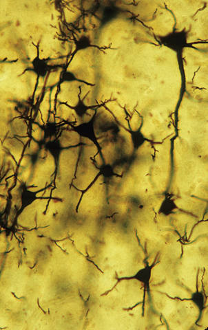
 Figure 3.1: Golgi-
Humans have thoughts, feelings, and behaviours that are often accompanied by visible signals. Consider how you might feel on your way to meet a good friend. An observer might see a smile on your face or notice how fast you are walking; internally, you might mentally rehearse what you will say to your friend and feel a surge of happiness as you approach her. All those visible and experiential signs are coordinated by the activity of your brain cells. The anticipation you have, the happiness you feel, and the speed of your feet are the result of information processing in your brain. All of your thoughts, feelings, and behaviours spring from cells in the brain that take in information and produce some kind of output trillions of times a day. These cells are neurons, cells in the nervous system that communicate with one another to perform information-
3.1.1 Components of the Neuron
During the 1800s, scientists began to turn their attention from studying the mechanics of limbs, lungs, and livers to studying the harder-
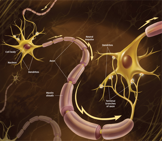
Cajal discovered that neurons are complex structures composed of three basic parts: the cell body, the dendrites, and the axon (see FIGURE 3.2). Like cells in all organs of the body, neurons have a cell body (also called the soma), the largest component of the neuron that coordinates the information-

 Figure 3.2: Components of a Neuron A neuron is made up of three parts: a cell body that houses the chromosomes with the organism’s DNA and maintains the health of the cell; dendrites that receive information from other neurons; and an axon that transmits information to other neurons, muscles, and glands.
Unlike other cells in the body, neurons have two types of specialized extensions of the cell membrane that allow them to communicate: dendrites and axons. Dendrites receive information from other neurons and relay it to the cell body. The term dendrite comes from the Greek word for “tree”; indeed, most neurons have many dendrites that look like tree branches. The axon carries information to other neurons, muscles, or glands. Axons can be very long, even stretching up to a metre from the base of the spinal cord down to the big toe.
Which components of a neuron allows it to communicate?
In many neurons, the axon is covered by a myelin sheath, an insulating layer of fatty material. The myelin sheath is composed of glial cells (named for the Greek word for “glue”), which are support cells found in the nervous system. Although there are 100 billion neurons busily processing information in your brain, there are 10 to 50 times that many glial cells serving a variety of functions. Some glial cells digest parts of dead neurons, others provide physical and nutritional support for neurons, and others form myelin to insulate the axon. An axon insulated with myelin can more efficiently transmit signals to other neurons, organs, or muscles. In fact, with demyelinating diseases, such as multiple sclerosis, the myelin sheath deteriorates, slowing the transmission of information from one neuron to another (Schwartz & Westbrook, 2000). This leads to a variety of problems, including loss of feeling in the limbs, partial blindness, and difficulties in coordinated movement and cognition (Butler, Corboy, & Filley, 2009).
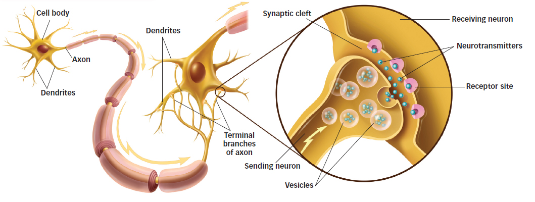
Cajal also observed that the dendrites and axons of neurons do not actually touch each other. There is a small gap or cleft between the axon of one neuron and the dendrites or cell body of another. This gap is part of the synapse, the junction or region between the axon of one neuron and the dendrites or cell body of another (see FIGURE 3.3). Many of the 100 billion neurons in your brain have a few thousand synaptic junctions, so it should come as no shock that most adults have 100 to 500 trillion synapses. As you will read shortly, the transmission of information across the synapse is fundamental to communication between neurons, a process that allows us to think, feel, and behave.

 Figure 3.3: The Synapse The synapse is the junction between the dendrites of one neuron and the axon or cell body of another. Notice that neurons do not actually touch one another: There is a small synaptic space between them across which information is transmitted.
3.1.2 Major Types of Neurons
How do the three types of neurons work together to transmit information?
There are three major types of neurons, each performing a distinct function: sensory neurons, motor neurons, and interneurons. Sensory neurons receive information from the external world and convey this information to the brain. They have receptors on their dendrites that receive signals for light, sound, touch, taste, and smell. For example, sensory neurons’ endings in our eyes are sensitive to light. Motor neurons carry signals from the spinal cord to the muscles to produce movement. These neurons often have long axons that can stretch to muscles at our extremities. However, most of the nervous system is composed of the third type of neuron, interneurons, which connect sensory neurons, motor neurons, or other interneurons. Some interneurons carry information from sensory neurons into the nervous system, others carry information from the nervous system to motor neurons, and still others perform a variety of information-
3.1.3 Neurons Specialized by Location
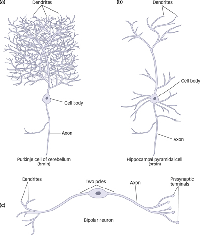
Besides specialization for sensory, motor, or connective functions, neurons are also somewhat specialized depending on their location (see FIGURE 3.4). For example, Purkinje cells are a type of interneuron that carries information from the cerebellum to the rest of the brain and spinal cord. These neurons have dense, elaborate dendrites that resemble bushes. Pyramidal cells, found in the cerebral cortex, have a triangular cell body and a single, long dendrite among many smaller dendrites. Bipolar cells, a type of sensory neuron found in the retinas of the eyes, have a single axon and small dendrites that feed into a single long dendrite that connects to the cell body. The brain processes different types of information, so a substantial amount of specialization at the cellular level has evolved to handle these tasks.

 Figure 3.4: Types of Neurons Neurons have a cell body, an axon, and at least one dendrite. The size and shape of neurons vary considerably, however. The Purkinje cell has an elaborate treelike assemblage of dendrites (a). Pyramidal cells have a triangular cell body and a single, long dendrite with many smaller dendrites (b). Bipolar cells have only one dendrite and a single axon (c).
Neurons are the building blocks of the nervous system: They process information received from the outside world, communicate with one another, and send messages to the body’s muscles and organs.
Neurons are composed of three major parts: the cell body, the dendrites, and the axon.
The cell body contains the nucleus, which houses the organism’s genetic material.
Dendrites receive sensory signals from other neurons and transmit this information to the cell body.
Each axon carries signals from the cell body to other neurons or to muscles and organs in the body.
Neurons do not actually touch. They are separated by a small gap or cleft, which is part of the synapse across which signals are transmitted from one neuron to another.
Glial cells provide support for neurons, usually in the form of the myelin sheath, which coats the axon to facilitate the transmission of information. In demyelinating diseases, the myelin sheath deteriorates.
Neurons are differentiated according to the functions they perform. The three major types of neurons include sensory neurons (e.g., bipolar neurons), motor neurons, and interneurons (e.g., Purkinje cells).