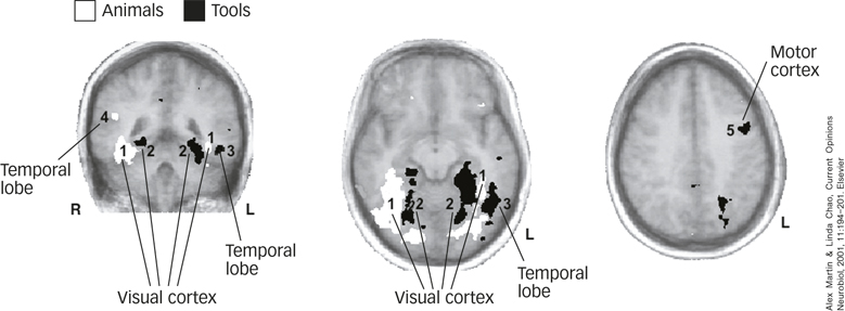
FIGURE 9.7 Brain Areas Involved in Category-Specific Processing Participants were asked to silently name pictures of animals and tools while they were scanned with fMRI. Areas in white, including parts of visual cortex and temporal lobe, labeled (1, 4), showed greater activity when participants named animals. Areas in black, including other parts of the visual cortex and temporal lobe (2, 3) and a region of the motor cortex (5), showed greater activity when participants named tools. Note that the images are left/right reversed.
Alex Martin & Linda Chao, Current Opinions Neurobiol, 2001, 11:194–201. Elsevier